
|
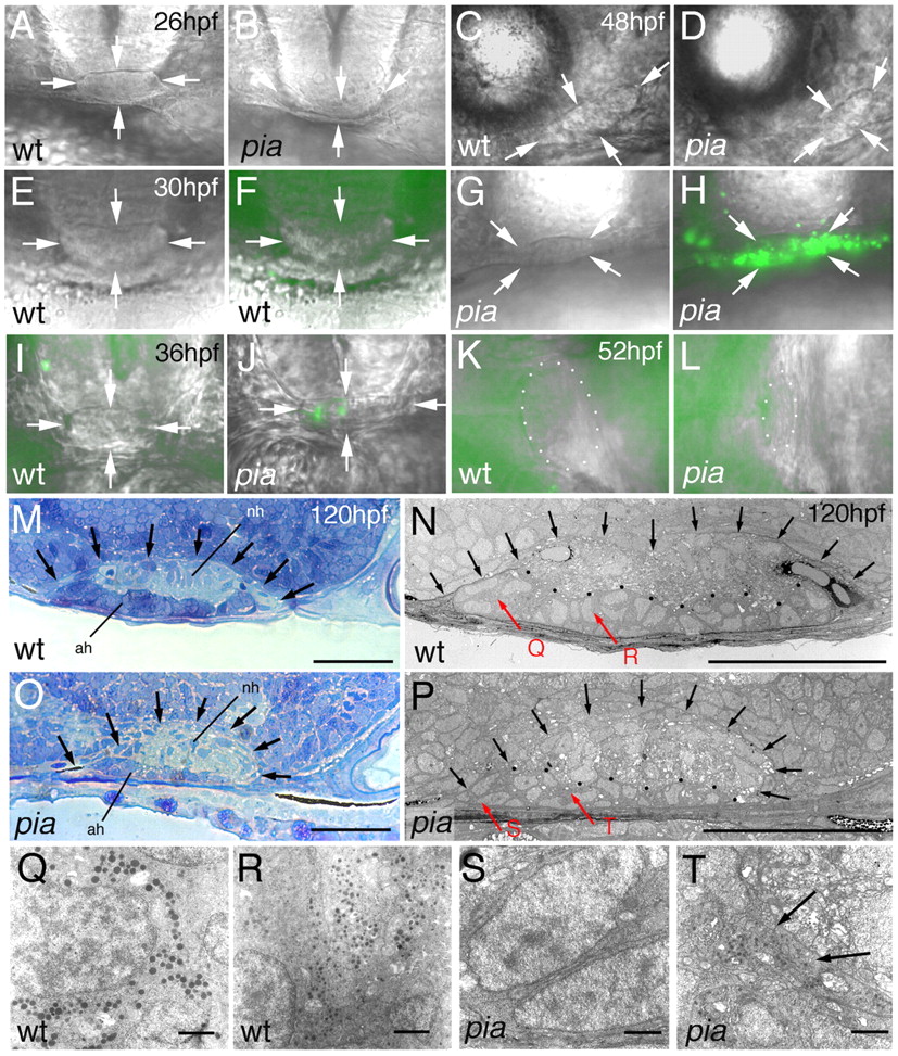
Fig. 4 pia mutants display indistinct early pituitary morphology, followed by a transient phase of adenohypophyseal cell death and the formation of a smaller, but distinct, pituitary gland with cells of rather primitive ultrastructure. (A-L) Nomarski images of live pia mutant embryos (pia) and their corresponding wild-type siblings (wt). (F,H,I-L) Images are superimposed by Acridine Orange staining for apoptosis. (A,B,E-J) Frontal views, dorsal upwards; (C,D) lateral views, anterior towards the left, dorsal upwards; (K,L) ventral views, anterior towards the right. Ages of embryos are indicated in upper right-hand corners of wild types. Genotypes were determined via PCR after photography. Arrowheads in A-J indicate borders of the pituitary gland; in K,L, pituitary borders are outlined by dots. (M-T) Pituitary ultrastructure at 120 hpf; longitudinal sections, anterior towards the left, dorsal towards the top. (M,O) Toluidine Blue staining; (N,P-T) electron micrographs. (M-P) The border of the pituitary is indicated by arrows; (N,P) the border between adenohypophysis (ah) and neurohypohysis (nh) is outlined by black dots. (Q-T) Higher magnifications of regions indicated by red arrows in N,P. Vesicles (as in T, indicated by arrows) were seen in three out of ~20 adenohypophyseal cells present in the section of the pia pituitary (P). They could contain matrix proteins and hormone-binding proteins, which can be made even in the absence of hormone production (compare with Norris, 1997). Scale bars: 30 μm in M-P; 1 μm in Q-T.