
|
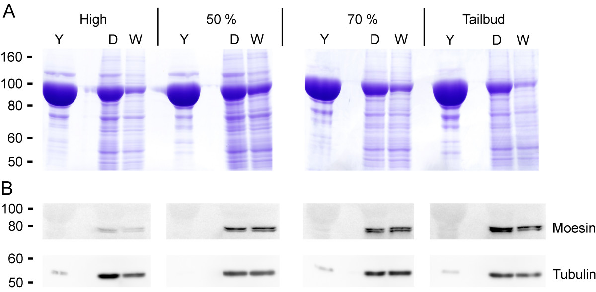
Fig. 3 Improved Western blotting results. Embryos at high stage (3 1/3 hpf), 50% epiboly (5 1/4 hpf), 70% epiboly (7 hpf) and tailbud stage (10 hpf) were deyolked, separated by SDS-gel electrophoresis and Coomassie stained (A) or blotted and immunodetected with antibodies against Tubulin (55 kD) and Moesin (78/80 kD apparent molecular weight) (B). Note that total protein amount was lower in deyolked samples, therefore more embryos could be loaded per lane: 1 embryo with yolk (Y), 15 embryos deyolked (D), 15 embryos deyolked and washed twice (W). Consequently, signal intensities of cellular proteins were increased.