
|
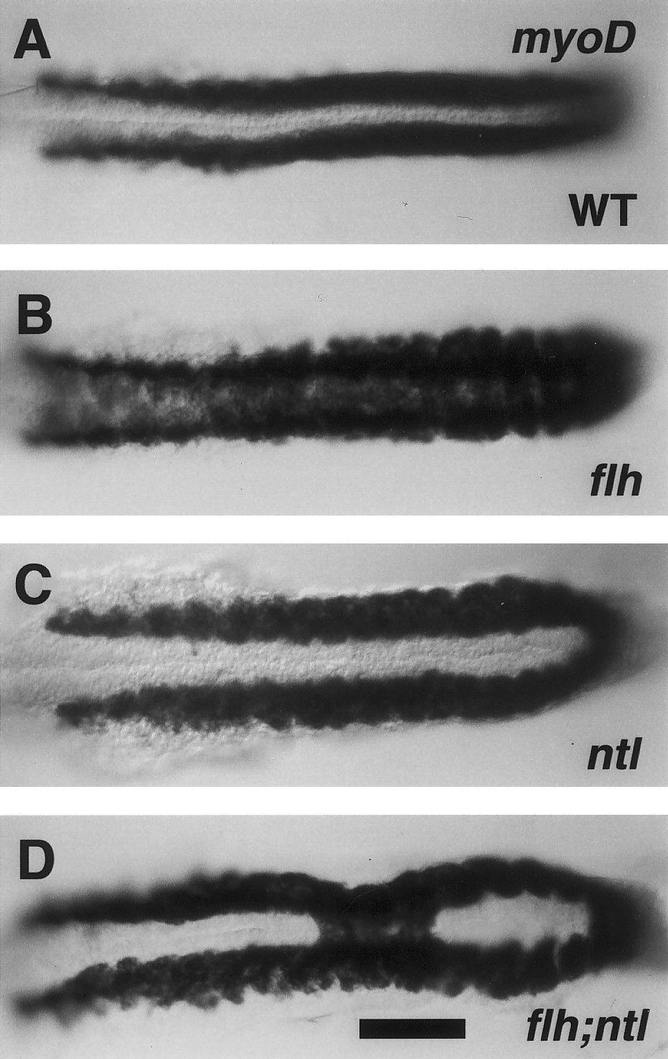
Fig. 5 ntl- partially suppresses somitic fusion due to flh-. Dorsal views, with anterior to the left of 19-hr embryos (20-somite stage). RNA in situ hybridization for myoD (Weinberg et al., 1996). In wild-type embryos (A), bilateral stripes of labeling are present in the trunk beside the developing notochord, which is unlabeled at the midline. In flh mutants (B) labeling spreads across the midline, whereas in ntl mutants (C) the stripes are distinctively farther apart than in the wild type (Odenthal et al., 1996). Labeling in the flh;ntl double mutant is generally ntl--like, but a single patch of tissue, involving three adjacent segments, shows the flh--like pattern. Forty-six percent of the double mutants (n = 13) had fusion of myoD expression that included regions of 2 – 5 somites (mean, 3.2 somites; n = 6) at midtrunk levels. The other flh;ntl double mutants were indisinguishable from ntl-. Genotyping of flh alleles was performed on all ntl mutant and flh;ntl mutant embryos by PCR analysis. Scale bar, 100 mm.
Reprinted from Developmental Biology, 187(2), Halpern, M.E., Hatta, K., Amacher, S.L., Talbot, W.S., Yan, Y.-L., Thisse, B., Thisse, C., Postlethwait, J.H., and Kimmel, C.B., Genetic interactions in zebrafish midline development, 154-170, Copyright (1997) with permission from Elsevier. Full text @ Dev. Biol.