
Fig. 4
- ID
- ZDB-IMAGE-060110-6
- Publication
- Lamason et al., 2005 - SLC24A5, a putative cation exchanger, affects pigmentation in zebrafish and humans
- All Figures
- Figures for Lamason et al., 2005
|
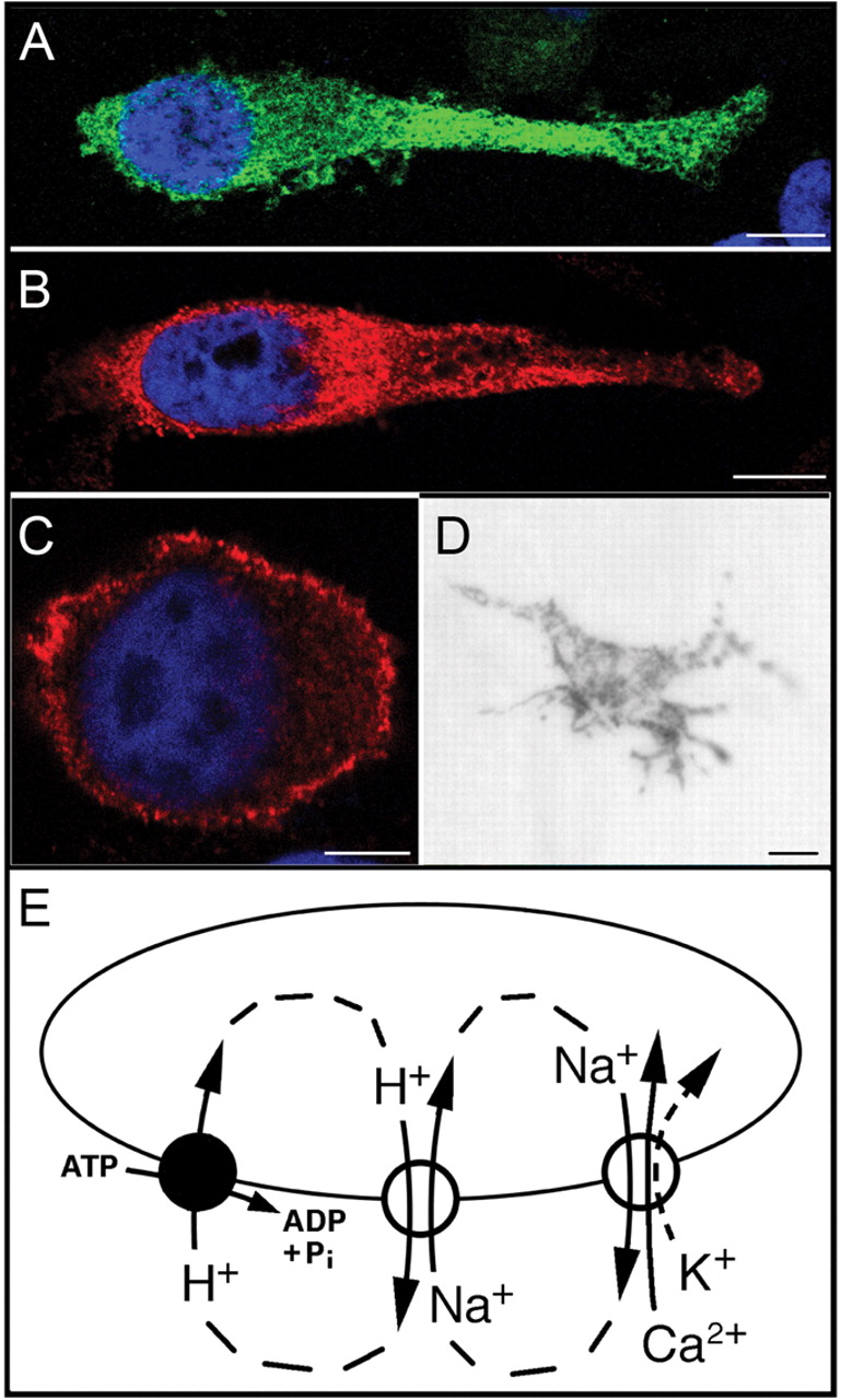
Fig. 4 Subcellular localization of slc24a5. Human MNT1 cells transfected with (A) GFP-tagged zebrafish slc24a5 (green) and (B) HA-tagged slc24a5 (red) clearly show intracellular expression. (C) HA-tagged D3 dopamine receptor localizes to the plasma membrane in MNT1 cells (red). 4′,6′-diamidino-2-phenylindole (DAPI) counterstain was used to visualize nuclei (blue). Scale bars in (A) and (B), 10 µm; in (C), 5 µm. (D) Rescue of dark pigmentation in a melanophore of a golden embryo by HA-tagged slc24a5. These dark cells appear in golden embryos injected with the HA-tagged construct, but not in mock-injected embryos. Scale bar, 10 µm. (E) Model for calcium accumulation in melanosomes. Protons are actively transported into the melanosome by the V-ATPase (left). The proton electrochemical potential gradient drives sodium uptake via the sodium (Na+)/proton (H+) exchanger (center). Sodium efflux is coupled to calcium uptake by the slc24a5 polypeptide (right). If potassium (dashed arrow) is cotransported with calcium, it must either accumulate within the melanosome or exit by means of additional transporters (not depicted). Pi, inorganic phosphate; ADP, adenosine diphosphate.