
Fig. 3
- ID
- ZDB-IMAGE-051214-3
- Publication
- Jin et al., 2005 - Cellular and molecular analyses of vascular tube and lumen formation in zebrafish
- All Figures
- Figures for Jin et al., 2005
|
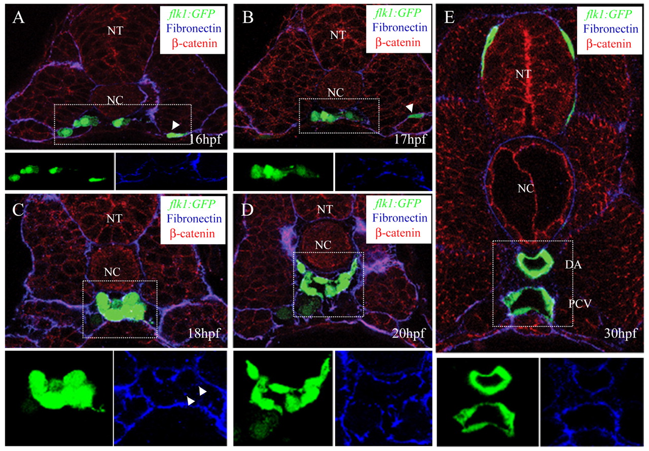
Fig. 3 Angioblast migration to the midline. (A-E) Transverse sections visualized for GFP (green), fibronectin (blue) and ß-catenin (red). The GFP (green) and fibronectin (blue) signals of the outlined areas are also shown separately. The sections shown are at the level of the 6th (A,B), 10th (C) and 14th (D,E) somite. White arrowheads in A and B mark angioblasts that are still residing within the LPM and that will migrate during a second wave. White arrowheads in C show fibronectin deposition around a single endothelial cell within the vascular cord. Over the span of 14 hours, the angioblasts migrate to the midline where they aggregate to form a vascular cord that subsequently lumenizes. NT, neural tube; NC, notochord; DA, dorsal aorta; PCV, posterior cardinal vein.