
Fig. 3
- ID
- ZDB-IMAGE-051107-2
- Genes
- Publication
- Honjo et al., 2005 - Slow muscle regulates the pattern of trunk neural crest migration in zebrafish
- All Figures
- Figures for Honjo et al., 2005
|
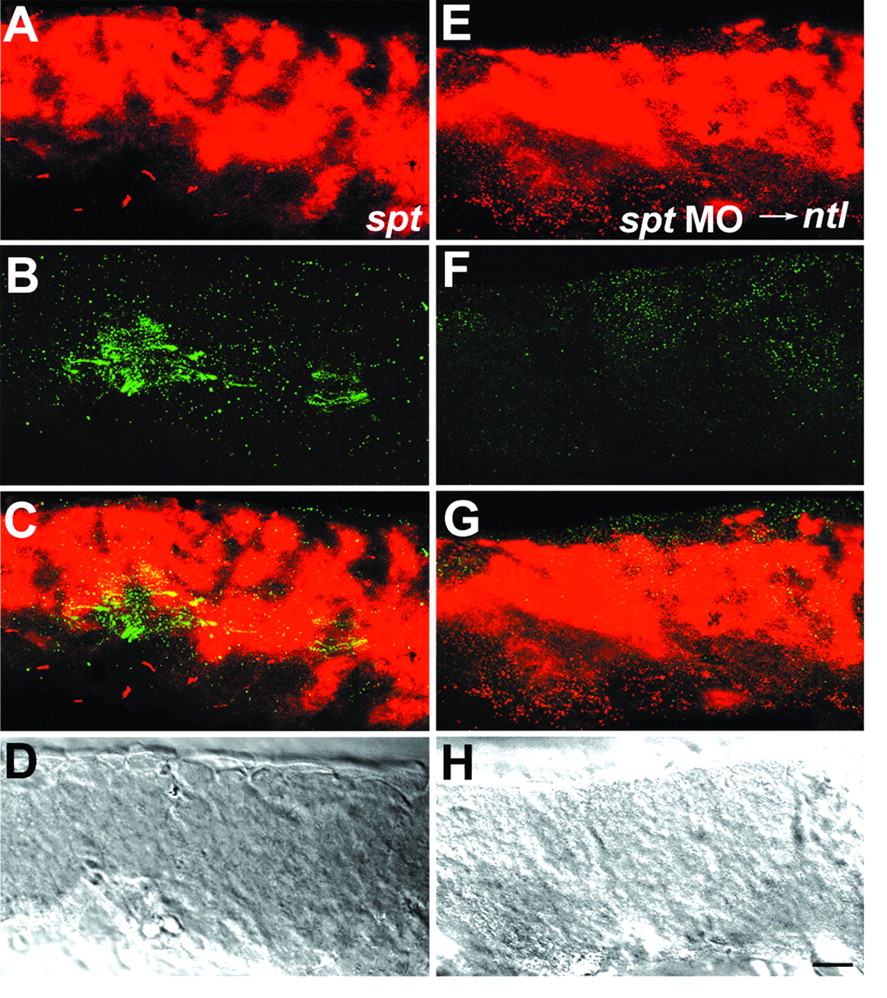
Fig. 3 Neural crest cells migrate in an unrestricted pattern in spt mutants and ntl mutants injected with spt MOs. (A-D) Whole-mount staining of 21 hpf spt mutant with crestin riboprobe (red) and F59 antibody (green). (A) Neural crest alone, (B) slow muscle alone, (C) the merged image, (D) a DIC image. F59 antibody staining reveals that spt mutants have few slow muscle cells. Neural crest cells migrate in spt mutants, but migration is not restricted to a specific pathway, thus there are no migration streams. (E-H) Whole-mount staining of 21 hpf ntl mutant injected with spt MOs. (E) Neural crest alone, (F) slow muscle alone, (G) the merged image, (H) a DIC image. F59 antibody staining reveals that ntl mutants injected with spt MOs have no muscle. As in spt mutants, neural crest migrates in these embryos, but migration is not restricted to a specific pathway and there are no migration streams in ntl mutants injected with spt MOs that have no muscle cells, as shown by absence of myod expression (H). Scale bar: 20 µm.