
Fig. 2
- ID
- ZDB-IMAGE-051107-1
- Genes
- Publication
- Honjo et al., 2005 - Slow muscle regulates the pattern of trunk neural crest migration in zebrafish
- All Figures
- Figures for Honjo et al., 2005
|
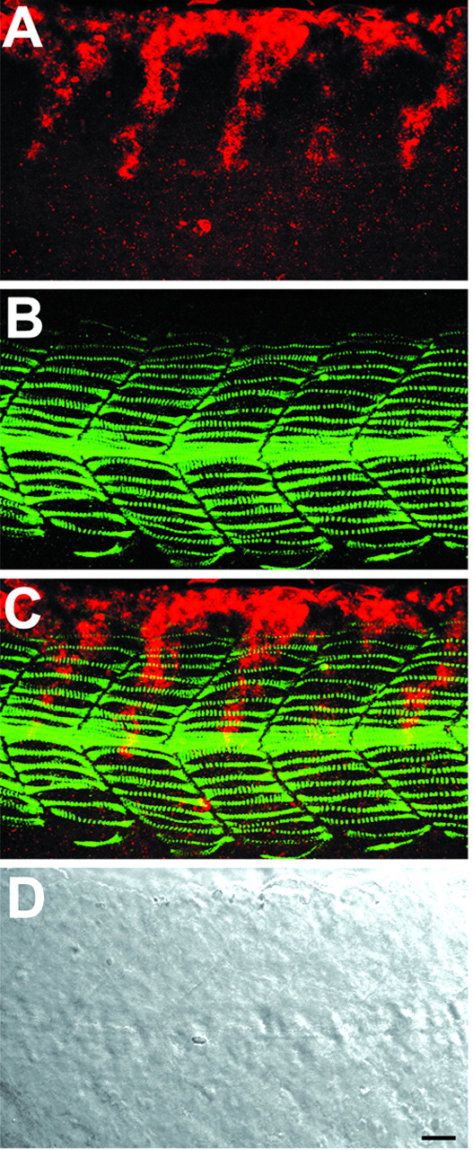
Fig. 2 Neural crest migration in wild-type embryos. Whole-mount staining of 21 hpf wild-type embryo with crestin riboprobe (red) and F59 antibody (green) to reveal trunk neural crest and slow muscle cells. (A) Neural crest alone, (B) slow muscle alone, (C) the merged image, (D) a Nomarski differential interference contrast (DIC) image. Neural crest migrates in the middle of the medial aspect of the myotome in one- to two-cell wide streams. Slow muscle fibers extend throughout the anteroposterior axis of each somite. At this stage of development, midtrunk level neural crest cells are in a more medial focal plane than slow muscle fibers, but they are seen together in this z-projection. Similar z-projections are shown in subsequent figures. Lateral view, anterior toward the left. Scale bar: 20 µm.