
Fig. 10
- ID
- ZDB-IMAGE-050930-16
- Publication
- Babb et al., 2005 - Zebrafish R-cadherin (Cdh4) controls visual system development and differentiation
- All Figures
- Figures for Babb et al., 2005
|
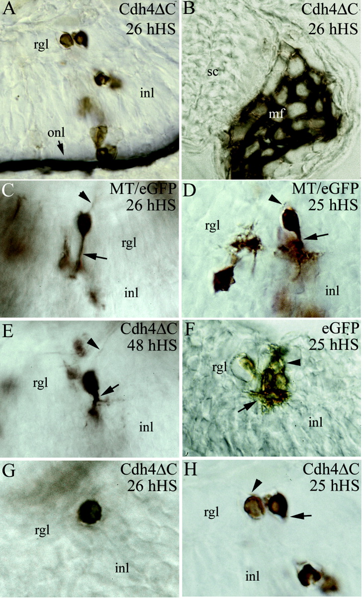
Fig. 10 Retinal ganglion cells (RGCs) expressing Cdh4ΔC at 24-26 hours postfertilization (hpf) have few or no processes at 56 hpf. A,B,D,F,H: Photomicrographs from retinal sections. C,E,G: Photomicrographs from whole-mount embryos. A,B: Shown are Cdh4ΔC-expressing cells in the retina (A) and in trunk skeletal muscle fibers (B) that were heat shocked at 25-26 hpf. A: A cross-section of the eye (dorsal to the right and lateral above) showing Cdh4ΔC-expressing cells throughout the thickness of the retina. B: A cross-section (dorsal above and lateral to the right) of the body trunk region showing that the anti-myc immunostaining was located mainly in the periphery of each muscle fibers (mf). C,D,F: Control RGCs expressing either MT/eGFP (C,D) or eGFP (F). These RGCs have numerous dendrites (arrow) and each has an axon (arrowhead). A,G,H: RGCs expressing the dominant-negative Cdh4ΔC (heat shocked at 25-26 hpf) have many fewer or no processes. E: A Cdh4ΔC-expressing RGC (heat shocked at 48 hpf) with normal RGC morphology. gcl, retinal ganglion cell layer; hHS, hours heat shock; inl, inner nuclear layer; onl, outer nuclear layer; sc, spinal cord; eGFP, enhanced green fluorescent protein.