
Fig. 8
- ID
- ZDB-IMAGE-050930-14
- Publication
- Babb et al., 2005 - Zebrafish R-cadherin (Cdh4) controls visual system development and differentiation
- All Figures
- Figures for Babb et al., 2005
|
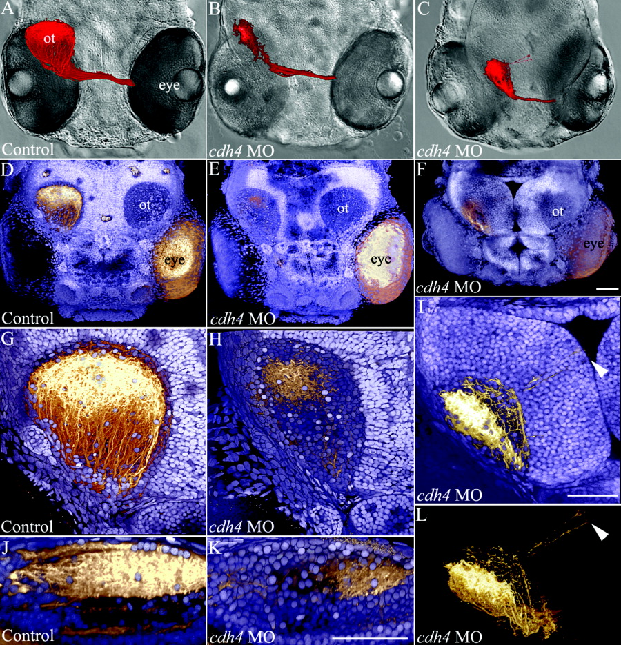
Fig. 8 Retinal ganglion cell axons in 5 days postfertilization (dpf) cdh4 knockdowns project to the contralateral optic tectum but do not arborize normally. A-L: Zebrafish embryos were injected with either standard control morpholino oligonucleotide (MO, A,D,G,J) or cdh4 MO (B,C,E,F,H,I,K,L). Embryos were fixed at 5 dpf and retinotectal projections were traced by injecting one eye in each embryo with the lipophilic dye DiI (1,1′-dioctadecyl-3,3,3′,3′-tetramethylindocarbocyanine perchlorate). Projection images of two-photon volumes of the DiI-filled retinotectal projections (shown in red) were overlaid with differential interference contrast (DIC) images from the same embryo (A-C). Staining within the injected eye was digitally removed to leave the DIC image of the eye visible. A-C: In controls (A), retinal ganglion cells project contralaterally and arborize to fill the optic tectum. In cdh4 knockdowns (B,C), retinal ganglion cells project to the contralateral optic tectum but do not arborize normally. D-L: These same three fish are shown at higher resolution. A-C: The DiI-injected fish were counterstained with DAPI (4′,6-diamidine-2-phenylidole-dihydrochloride) so that the boundaries of the tectal neuropil could be seen in relationship to the retinotectal projections (shown in gold). DAPI (shown blue-violet) stains the cell nuclei. Cellular regions are filled with nuclei, but nuclei are absent from regions of neuropil such as the optic tectum (ot). D,G,J: In controls (D,G,J) from a dorsal view (D,G), the retinotectal projections arborized to fill the entire optic tectum. J: A medial view of a sagittal cut through the optic tectum of the control volume shows the deep tectal layers. E,F,H,I,K,L: In cdh4 knockdowns, both retinotectal projections and the optic tectum were affected. E,H: Optic tecta in some moderately affected cdh4 knockdowns were apparently normal in size, but the retinotectal projections were sparse, disorganized, and did not fill the tectal neuropil. K: A medial view of a sagittal cut through the optic tectum of the moderately affected cdh4 knockdown lacks the well defined deep layers. F,I,L: In severe knockdowns, the tectal neuropil was smaller (F,I) and some neurites extended beyond the neuropil boundary and into the cellular layer (arrowheads I,L). Scale bars = 50 μm.