
|
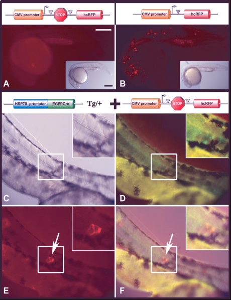
Fig. 5 Detection of hcRFP after heat shock induction of EGFP-Cre. A: Lack of detectable expression from the hcRFP reporter construct (depicted at the top of the panel) in mosaic transgenic embryos at 24 hours postfertilization (hpf). B: Detection of expression from final recombination product (depicted at the top of the panel) in mosaic transgenic embryos at 24 hpf. C-F: Detection of hcRFP-positive cells after heat shock induction of EGFP-Cre. C: Brightfield image showing the trunk of an HSP70-EGFP-cre embryo injected with the hcRFP reporter construct. D: EGFP fluorescent image of embryo shown in C, showing remaining EGFP intensity 24 hr post heat shock. E: hcRFP fluorescent image of embryo shown in C. Arrow denotes a cluster of hcRFP-positive cells 24 hr post heat shock. F: Artificial merge of brightfield and fluorescent images. Arrow denotes the cluster of hcRFP-positive cells shown in E. Scale bars = 250 microns in A (applies to A,B and insets).