
|
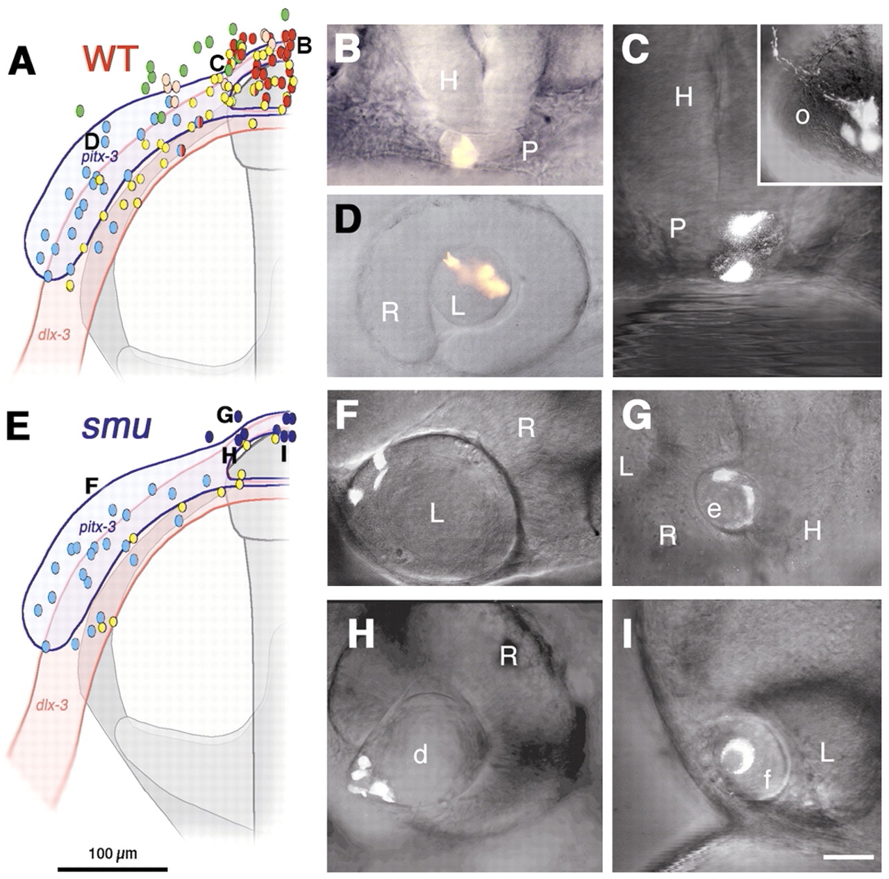
Fig. 3 Pituitary precursors form lens in smoothened mutant embryos. (A-D) Wild-type embryos, (E-I) smu mutant embryos. Colored dots and letters in A,E represent locations of injected precursor cells at bud stage and their fates at prim-5 stage (B-D,F-I). (A) Fate map of wild-type embryo (n=105) shows pituitary precursor cells (red) at anterior midline (B, n=22), lens precursor cells (blue) at lateral neural plate border (D, n=21) and olfactory placode precursors (yellow) closer to neural plate (C, inset, n=37). (C) Split fates from a single precursor cell (yellow/red dots in A) contribute to pituitary and olfactory placode. Green dots, epidermal precursors; pink dots, head mesenchymal precursors. (E) Fate map of smoothened mutant embryo (n=33). Precursor cells give rise to ectopic median (dark blue) or normal retinal lenses (light blue); yellow, olfactory placode precursors (n=9). (F) Precursor cells from lateral pitx3 domain (light blue) give rise to lens epithelial and primary lens fiber cells. (G-I) Median placodal precursors give rise to ectopic (G), distorted (H) or fused (I) lens, and not to pituitary as expected from median positions in wild-type embryos (A, red). Ventral ectoderm and head mesenchyme cells not shown for clarity. d, distorted lens; e, ectopic lens; f, medially fused lens; H, hypothalamus; L, retina associated lens; o, olfactory placode; P, pituitary; R, retina. (A,B) Dorsal view, prospective head, anterior towards the top (compare with Fig. 2I). (B,C,G) Frontal view, dorsal towards the top. (D,F,H,I) Side view, anterior towards the left. Scale bar: 100 μm in A,E; 50 μm in B,D,E; 25 μm in C,F-I.