
|
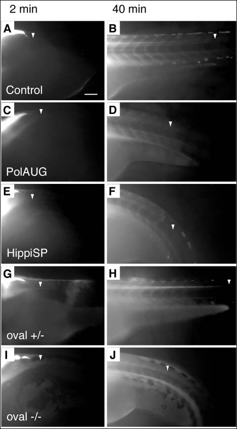
Fig. 5 Fluid flow is impaired by lack of normal cilia movement in the central canal of the spinal cord. BDM-pretreated embryos were injected with 5% tetramethylrhodamine-conjugated 70 k MW dextran into the fourth brain ventricle and dye distribution along the central canal of the spinal cord was recorded at various timepoints; shown are 2 and 40 minutes post-injection. Control (A,B) and oval heterozygous (G,H) show a distribution of the dye up to an anterior-posterior level of the tip of the yolk extension at 40 minutes (arrowheads) (B,H), whereas polarisAUG morphant (C,D), hippiSP morphant (E,F) and oval homozygotes (I,J) show reduced dye migration. Anterior to the left. Scale bar: 100 µm.