
|
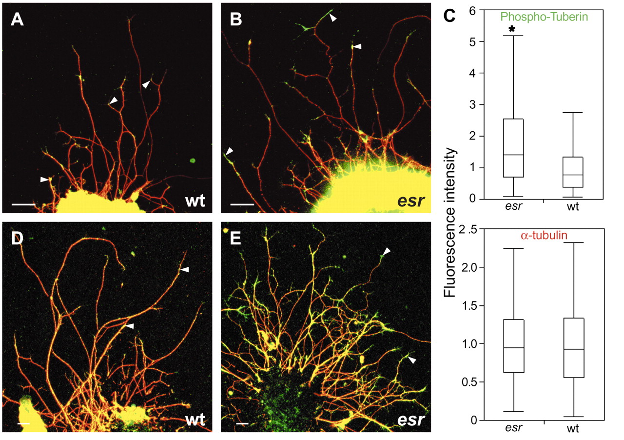
Fig. 7 Ser939 Phospho-Tuberin (green) and -tubulin (red) immunofluorescence in retinal explants. (A) After 15 hours in culture, Phospho-Tuberin (arrowheads) is detected in retinal axons, with higher levels in the esrom mutant (B). (C) At this time, Phospho-Tuberin immunofluorescence is twofold higher on average in esrom axon tips (*P<0.0001); box plot shows medians and quartiles from two normalized independent experiments (n wt=83, n esr=99 and n wt=26, n esr=78). After 26 hours in culture, the fasciculation phenotype is apparent, and Phospho-Tuberin staining difference is intensified. Scale bar: 50 µm