adult zebrafish; drawings and H&E staining structure of the mature testis - tubules
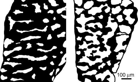
The zebrafish testis is composed of anastomosing seminiferous tubules. In these images, the tubule lumina are highlighted (white) to enhance the testis structure; the masked image
 and the unmasked image
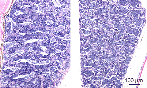 are given for comparison.
The left specimen shows that the zebrafish testis has a tubular structure; the right-hand specimen is sectioned differently, thus showing its anastomosing character - branching tubules are folded and interwoven to form the organ, in line with the characterization of teleost testis by Grier
are given for comparison.
The left specimen shows that the zebrafish testis has a tubular structure; the right-hand specimen is sectioned differently, thus showing its anastomosing character - branching tubules are folded and interwoven to form the organ, in line with the characterization of teleost testis by Grier 1,2.
1,2.

adult zebrafish; H&E staining structure of the mature testis - tubules
Each tubule is bounded by a basement membrane and a connective tissue sheet
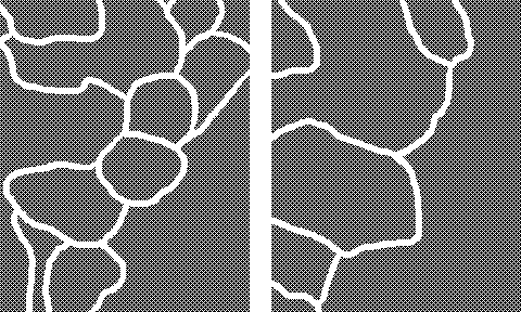 (see details). Most tubules are covered with seminiferous epithelium
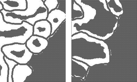 , the structure of which is shown on the spermatocysts page.
Mature sperm is voided into the tubular lumen, which than serves as a storage compartment. The tubular lumen system is conntected to main efferent tubules (ducts
 ), which are located at the periphery of the organ (i.e.rostral and caudal poles, and the against median border, as shown here); these efferent tubules have no or only small clusters of seminiferous tissue. These areas should be avoided when analyzing the status of the testis.
References
- Grier-HJ, Linton-JR, Leatherland-JF, De Vlaming-VL. Structural evidence for two different testicular types in teleost fishes. Am. J. Anat. 159: 331-345; 1980.
- Grier-HJ. Comparative organization of Sertoli cells including the Sertoli cell barrier. In: Russell-LD and Griswold-MD (eds.), The Sertoli Cell: 703-739. Clearwater FL, Cache River Press 1993.