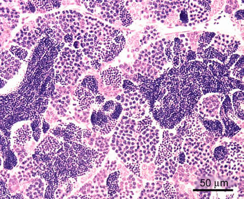
adult zebrafish; H&E staining structure of the mature testis - the seminiferous tubules are lined with cysts
The structural organisation of tubules
 is visible at medium power magnification.
In the image below it is also evident, that the lining seminiferous tissue is built of clusters of cells, the so called spermatocysts
 .
.The composition of these spermatocysts is best viewed at high magnification, see next section. The lumen
 of the tubules is filled with mature sperm.
Note, that zebrafish have continuous reproduction, and therefore show a balanced presence of proliferating, differentiating, maturing spermatogenic stages, and mature sperm. In contrast, the testis of seasonal breeders shows predominance of one of these successive stages predominates; these testis may be classified accordingly.