
Coronal section of a juvenile male zebrafish, 15 mm (6 weeks); H&E staining testis location, coronal view
The testis
 is a paired organ, located bilaterally between the abdominal wall [AW] and the swim bladder
 . At the level of this section, the testis is further bounded by the liver
 and mesenchymal tissue
  (mainly adipose tissue); dorsally it is adjacent to the kidney and ventrally to the intestines (not visible on this level).
This image further shows: the abdominal wall musculature
 , the head kidney
 , the opercles and gills
 , the pharyngeal cavity and epithelium
 , and the pharyngeal musculature
 .
.

Axial section of an adult male zebrafish; H&E staining testis location, axial view
This axial section of an adult male zebrafish is from the rostral part of the abdominal cavity, where the testis
 is in the upper part, between swim bladder
 and abdominal wall. The area of testis will show largely similar in more caudal sections.
The abdominal organs shown in this image are liver
 , pancreas
 , spleen
 , intestinal loops
 .
.
Major blood vessels in this section are dorsal aorta
 and posterior cardinal vein
 , embedded in the kidney
 . Intestinal arteries
 , intestinal veins
 and epigastric vein
 , partly embedded in the liver
 .
.
The horizontal skeletogenous septum
 separates the epaxialis
 and hypaxialis muscles
 , which are inserted to this septum and to the spine
  , and which are also bound to the skin. In the midline, the epaxial muscles
 insert to the vertical skeletogonous septum
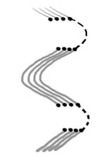
 . Although organized in myotomes which are oriented perpendicular to the length axis of the fish, these muscles appear cross-sectioned due to their waved course
 . The hypaxialis muscles
 includes the dorsal supracarinalis muscles
 , the hypaxialis muscles
 include the ventral infracarinalis muscles
 . The lateralis superficialis muscles
 overlay the peripheral part of the horizontal septum
 . Pleural rib structures
 can be observed within the hypaxialis muscles
 .
.