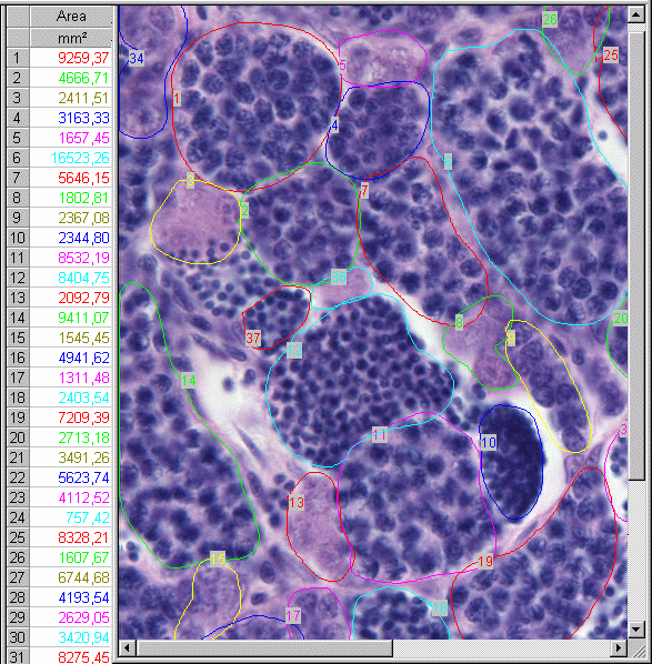
adult male zebrafish - mature testis mature testis This image shows part of a screenshot of the used image analysis package (AnalySIS, Soft Imaging System GmbH, Münster, Germany; other (free) programs (e.g. ImageJ) can do the same job). Digital images were loaded into the package, magnification calibrated and adjusted, and testis structures outlined. The package generates a table with figures corresponding to the outlined areas (shown in the left column, x10E7). Each figure was identified with a histological classifier, using three main stages of spermatogenesis: spermatogonium, spermatocyte, and spermatid. Thus the substages spermatogonium A and B, and the substages leptotene, zygotene and pachytene spermatocytes were pooled, as transition of spermatogium to spermatocyte, i.e. initiation of meiosis, and transition of spermatocyte to spermatid, i.e. initiation of final sperm maturation, were considered critical steps, sensitive to endocrine disruption.
Further calculations (total area and number for each class, average and relative sizes and numbers) were done in a spreadsheet.
Since the area of interest lays in the upper segment of previtellogenic maturation, a cut-off level of 0.0015 mm² was set to exclude an overrepresentation of the smallest development stages; the measurement was also not biased by vitellogenic oocytes, which grow faster than previtellogenic oocytes, because these were not present in the analysed ovaries.
Reference
- van der Ven-LT, Wester-PW, Vos-JG. Histopathology as a tool for the evaluation of endocrine disruption in zebrafish (Danio rerio). Environ. Toxicol. Chem. 2003;22:908-913.