
Coronal section of a juvenile male zebrafish, total body length 17 mm; age 8w; H&E staining testis-ova
Early oocytes
 are observed in the developing testis at varying prevalences (1:350 in the RIVM zebrafish stock, a higher prevalence was reported in the stock of the Veterinary Faculty, Utrecht University; Raoul Kuiper, personal communication). This image shows the phenomenon at a relatively more advanced developmental stages, which includes mature sperm
 . There is an obvious clustered organisation (spermatocysts
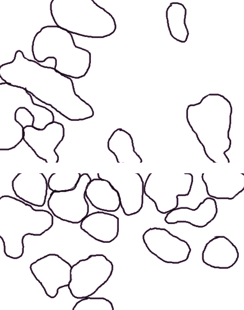 ), combined with clusters of spermatogonia
 . Incidently, Sertoli cells
 can be discerned.
The presence of primitive oocytes in the developing testis has been interpreted as a feature of undifferentiated gonochorism or protogyny: the undifferentiated gonad develops into an ovary-like gonad in all individuals and then in about one-half of the population transforms into a testis1. Takahashi described "intersexuality" similar to the images presented here with a maximal incidence of 1:4. These varying incidences indicate that the balance of sex-determining factors is delicate and environmental or genetical variations between stocks may produce a slightly different differentiation process. Our observations do not support the undifferentiated gonochoristic or protogyny paradigm in zebrafish.
Note: testis-ova is distinct from ovo-testis, where ovarian tissue is observed adjacent to testicular tissue.
Reference
1 Takahashi-H. Juvenile hermaphroditism in the zebrafish, Brachydanio rerio. Bull.Fac.Fish.Hokkaido Univ.28:57-65;1977