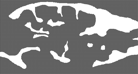
Coronal section of a juvenile male zebrafish, total body length 11 mm; age 6w; H&E staining 1. undifferentiated gonad
The undifferentiated gonad
 is an elongated structure which diametrically only spans a few cells; the organ is composed of mainly undifferentiated gonocytes. In this image, it is bounded by liver
  and its associated peritoneal tissue
  on the medial side, and lateral by mesenchyme
 . Adjacent to the liver, part of the swim bladder
 is visible.
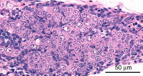
Coronal section of a juvenile male zebrafish, total body length 11 mm; age 4w; H&E staining 2. early development
An early feature of male differentiation of the gonad is ingrowth of stroma
 . Other features, which are not present on this image are appearance of a clustered organisation (spermatocysts) and lumen formation. This gonad mainly contains undifferentiated gonocytes
 , but early stages of differentiation
 are also present.
A small part of the liver
 is visible.

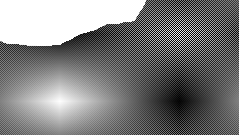
Coronal section of a juvenile male zebrafish, total body length 11 mm; age 5w; H&E staining 3. early development
This gonad mainly contains early stages of differentiation
 , which are characterized by the increased nuclear basophilic staining and decreased size, compared to undifferentiated gonocytes
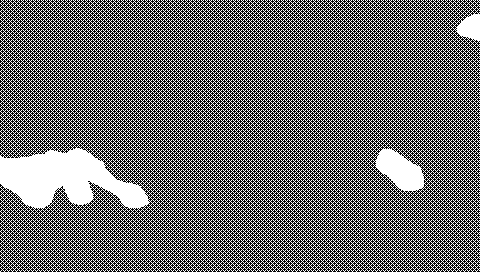 . The hole in this image probably indicates early lumen
 formation; early ingrowth of stroma
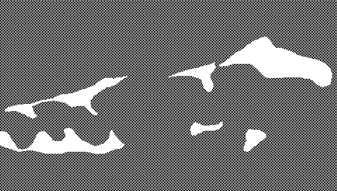 is also visible.
Adjacent structures in this image are: liver
 on the medial side and mesenchyme
 on the lateral side.

Coronal section of a juvenile male zebrafish, total body length 12 mm; age 5w; H&E staining 4. early development
Early features of male differentiation of the gonad are the appearance of a clustered organisation (spermatocysts)
 and ingrowth of stroma
 .
.This gonad
 contains early stages of differentiation
 , as well as undifferentiated gonocytes
 . A vessel
 can be observed, and adjacent structures in this image are: liver
 , pancreas
 , and peritoneal cells
 .
.
Coronal section of a juvenile male zebrafish, total body length 12 mm; age 5w; H&E staining 5. early development
This gonad shows the combined presence of clustered organisation (spermatocyst
 ) and lumen formation
 , as indications of early male differentiation.
Early stages of differentiation
 are predominant, but undifferentiated gonocytes
 are also abundant.
Adjacent structures in this image are: liver
 and eosinophilic peritoneal cells
 in mesenchyme.
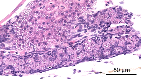
Coronal section of a juvenile male zebrafish, total body length 12 mm; age 6w; H&E staining 6. intermediate development
This gonad shows the combined presence of clustered organisation (spermatocysts
 ) and lumen formation
 as indications of early male differentiation. A prominent blood vessel
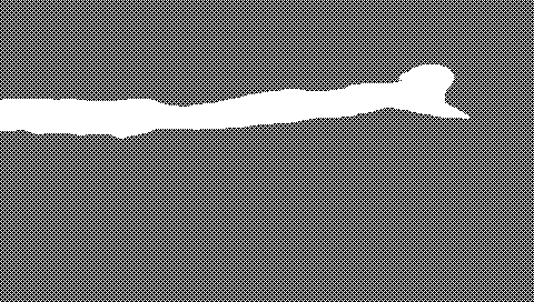 filled with erythrocytes
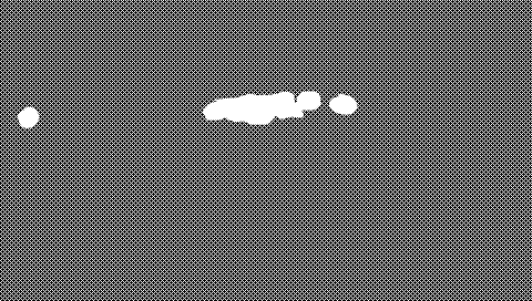 is present in the stroma.
The spematogenic epithelium contains mainly early and intermediate stages of differentiation
 , up to early spermatids
 ; clusters of undifferentiated gonocytes
 are also present.
Adjacent structures in this image are: liver
 and abdominal wall
 .
.
Coronal section of a juvenile male zebrafish, total body length 12 mm; age 6w; H&E staining 7. advanced development
This gonad can be identified as a testis by the presence of well-developed lumina
 , which are now filled with sperm, and by the clustered organisation (spermatocysts
 ). Clusters of spermatogonia
 are also present.
Adjacent structures in this image are: clustered peritoneal cells
 .
.