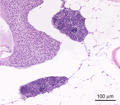
Total body length 14 mm; age 6w; H&E staining testis fully attached to the wall of the peritoneal cavity
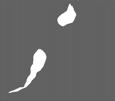
The differentiated gonad (testis in this image)
 develops from an undifferentiated gonadal anlage, located retroperitoneally in the caudal peritoneal cavity
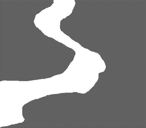 . It is closely associated with the peritoneum
 , from which ingrowth of stroma into the gonad originates. At this stage, the testis is in close association with the wall of the peritoneal cavity, and bounded by retroperitoneal mesenchymal tissue
 (mainly adipose cells).
 This image further shows part of the liver
 and of the swim bladder
 .
.

Total body length 12 mm; age 6w; H&E staining central detachment
Central detachment of the gonad
 of the peritoneal wall leads to the development of a retrogonadal cavity
 . At this stage, the gonad (testis in this image) remains attached
 to the peritoneal wall on both sides, and these attachments are continuous with the peritoneum
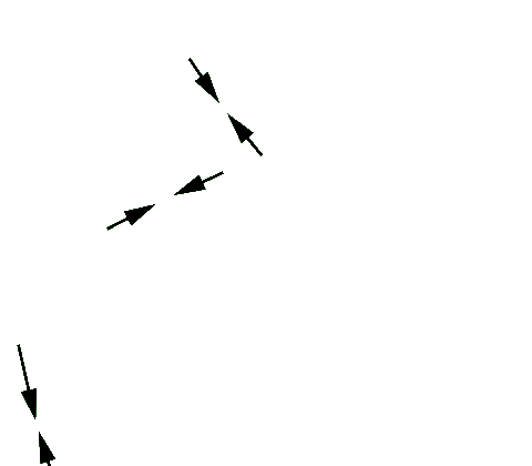 . The gonad now bulges more prominent into the peritoneal cavity
 ; it is no longer closely associated with the retroperitoneal mesenchyme
 .
.
This image further shows part of the intestine
 , pancreas
 , trunk wall muscles
 (which clearly shows somite organization
 and a cross-cut bone
 ), and of the swim bladder
 .
.
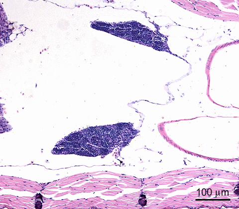
total body length 12 mm; age 6w; H&E staining unilateral attachment
After further detachment, the testis
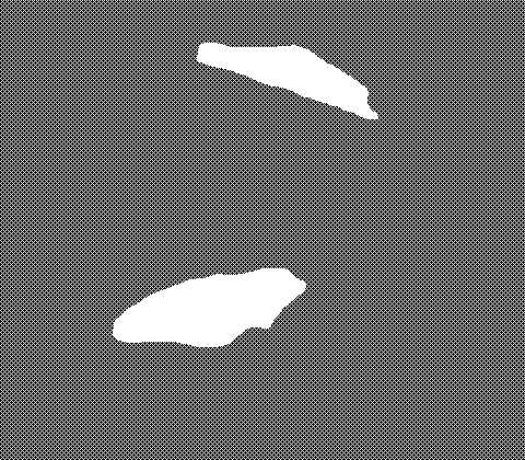 appears as a protruding mass in the peritoneal cavity
 . A single pole of the organ remains attached to the peritoneal wall
 . This attachment area, which is continuous with the peritoneum
 eventually develops to a testicular stalk, conducting the efferent sperm duct.
This image further shows part of the intestine
 , liver
 , trunk wall muscles
 (which clearly shows somite organization
 and a cross-cut bone
 ), of the retroperitoneal mesenchyme
 , and of the swim bladder
 .
.

total body length 13 mm; age 6w; H&E staining persisting bilateral attachement in the ovary
In ovary development, the retrogonadal cavity
 persists and even expands; eventually it becomes the oviduct. The ovary
 thus remains attached bilaterally to the peritoneal wall. The peritoneum
 constitues the boundary between this future oviduct and the peritoneal cavity
 .
.
This image further shows part of the intestine
 , liver
 , pancreas
 , trunk wall muscles
 (which clearly shows somite organization
 and a cross-cut bone
 ), of the retroperitoneal mesenchyme
 , and of the swim bladder
 .
.
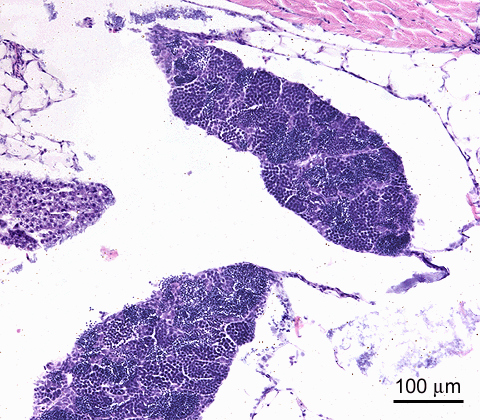
total body length 17 mm; age 6w; H&E staining persisting bilateral attachement in the testis
As shown in this image, the retrogonadal cavity
 may also persist in testis development. The testis
 thus remains attached bilaterally to the peritoneal wall
 . It should be noted, however, that this bilateral attachment may indicate feminization, since this animal was staticly exposed to 1 nM 17ß-estradiol (lifetime). The peritoneum
 constitues the boundary between this cavity and the peritoneal cavity
 .
.
This image further shows part of the liver
 , trunk wall muscles
 and of the retroperitoneal mesenchyme
 .
.