
adult male zebrafish exposed to 17a-methyl-dihydrotestosterone (MDHT) lower image; reference in upper image After exposure of adult zebrafish to the non-aromatisable androgen 17a-methyldihydrotestosterone (MDHT) for three weeks, the most striking effect in the testis is expansion of the interstitial compartment
 . On close examination, this expansion seems mainly the result of accumulation of proteinaceous intra- and extravascular fluid
 . This protein is probably overexpressed vitellogenin, as indicated by the basophilic staining of hepatocytes (not shown), similar to hepatic basophilia after estrogenic stimulation. This phenomenon suggests interaction of MDHT or one of its metabolites with the hepatic estrogen receptor. Pseudo-estrogenic effects are even more pronounced at higher concentrations (see below).
Visual inspection at low and medium power magnification reveals no further aberrations; in particular, there is no obvious imbalance in size and ratio of the main spermatogonic classes, i.e. spermatogonia
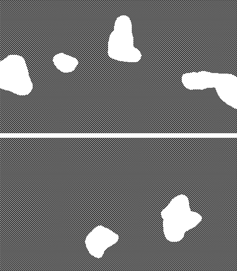 , spermatocytes
 , and spermatids
 . Lumen of the testis tubules is also normally filled with mature sperm. However, morphometrical analysis suggests that this androgenic stimulation induced an acceleration of spermatogenic maturation (see below).
Furthermore, at high power magnification, Sertoli cells are more easily discernible than in control testis (see below), suggesting hyperplasia or hyperproliferation of these cells.
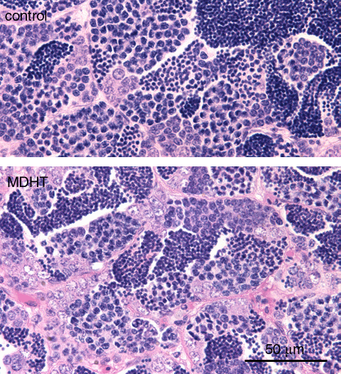
adult male zebrafish exposed to 17a-methyl-dihydrotestosterone (MDHT) lower image; reference in upper image After exposure to a high dose of the non-aromatisable androgen (1000 mg/l 17a-methyldihydrotestosterone; MDHT) for 10 days (lower image), the testis of adult male zebrafish showed increased size of the interstitial tissue compartment
 ; this effect is also observed in other tissues, and it is mainly due to vascular dilatation and interstitial accumulation of fluid, which in turn both are the result of overexpression of vitellogenin, as indicated by hepatic hyperbasophilia. This is reminiscent of effects induced after estrogenic exposure, and indicates interaction of MDHT or one of its metabolites with the hepatic estrogen receptor.
However, visual observation revealed no indication of changes in the ratio of spermatogenic stages

 , in contrast to the findings after estrogenic exposure.
 Morphometrical analysis (see below), on the other hand, showed that the ratio of the subsequent spermatogenic stages did change, towards the spermatid stage (in contrast to the shift towards the spermatogonium stage after exposure to estrogen).
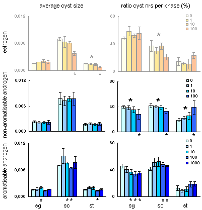
sg, spermatogonium; sc, spermatocytes; st, spermatids;
estrogen concentrations in nM/L; androgen concentrations in µg/L.
large *, significant dose-effect relation (univariate ANOVA);
small *, significant effect of a particular dose compared to control (T-test) The visual impression of relative changes of various spermatogenic stages is determined by two parameters: cyst size and relative number of cysts of these various stages in the field of view. Morphometrical analysis (see methods) of these two parameters after exposure to the non-aromatisable androgen 17a-methyldihydrotestosterone (MDHT) in the testis showed that the average cyst size is not at all influenced
 by MDHT. However, there is a change in the ratio of numbers of the respective cyst types, indicating a change in the rate of cyst development:
spermatogonium and spermatocyte cysts are relatively less frequently observed, and spermatid cysts more often
 , indicating that progression through these stages is accelerated by the androgen. Morphometry is required to discern this effect, since it was not observed on visual inspection (see above).
By contrast, the estrogen 17b-estradiol induces a decrease of spermatocyte and spermatid cyst sizes
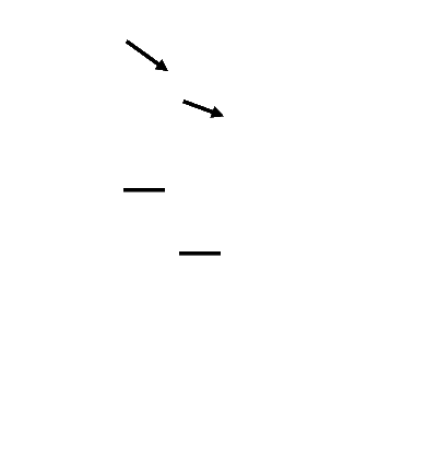 ; as with MDHT, E2 induces a decreased number of spermatocyte cysts, but in contrast to MDHT, exposure to E2 is accompanied by a constant or possibly even increased number of clusters of spermatogia
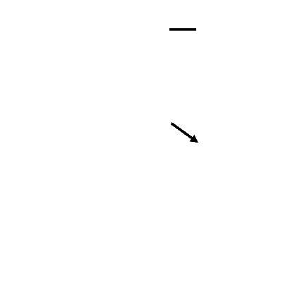 (the number of spermatid cysts possibly also increases after E2 exposure, as with MDHT).
The effects of the aromatisable androgen methyltestosteron are inconsistent, but in general more comparable to those of MDHT than of E2
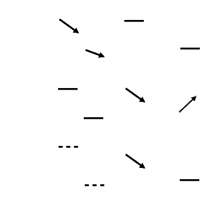 .
.

adult male zebrafish; top, control; middle and bottom: exposed to 1 mg/L 17a-methyldihydrotestosterone (MDHT) for 10 days After exposure of adult male zebrafish to the non-aromatisable androgen 17a-methyldihydrotestosterone (MDHT), Sertoli cells are more prominent
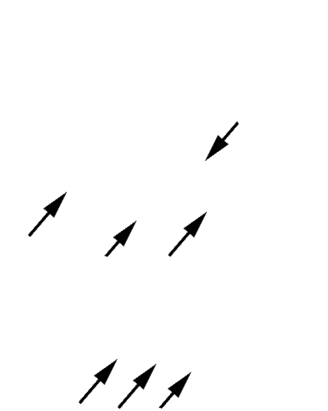 than in non-exposed specimen
 , due to nuclear hypertrophy. The clustered appearance of these cells in the bottom image suggests proliferation.