- Title
-
Mutations affecting development of the zebrafish inner ear and lateral line
- Authors
- Whitfield, T.T., Granato, M., van Eeden, F.J., Schach, U., Brand, M., Furutani-Seiki, M., Haffter, P., Hammerschmidt, M., Heisenberg, C.P., Jiang, Y.J., Kane, D.A., Kelsh, R.N., Mullins, M.C., Odenthal, J., and Nüsslein-Volhard, C.
- Source
- Full text @ Development
|
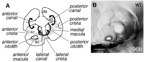
Structural features of the wild-type zebrafish ear. Anterior is to the left in all Figs. (A) Cut-away lateral view of a wild-type zebrafish ear at 96 hours, to give a three-dimensional impression of the structures within the vesicle. (B) Photograph of an ear at the same age, taken with differential interference contrast (DIC) optics, and focussed at the level of the anterior otolith. Note the overall shape of the organ. Epithelial projections (ep) within the ear form hubs of the developing semicircular canals (curved arrows). Each canal (anterior, lateral and posterior) is associated with a small sensory patch or crista, while each otolith overlies a larger sensory patch or macula. Kinocilia of the crista hair cells (kc) are long, and project into the canal lumens. Kinocilia of the maculae are shorter and the otoliths appear to sit directly on the stereociliary bundles (sc) of the macular hair cells. The smaller (anterior) otolith lies in a lateral position; the larger (posterior) otolith lies medially. dls, dorsolateral septum. Scale bar, 50 μm. |
|
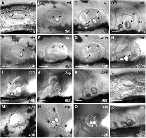
DIC images showing the ear phenotypes of live mutants. (A-D) Lateral views of wild-type (wt) ears, for comparison with the mutant phenotypes. Ages are given in hours (h) for each image. From 72-120 hours, the appearance of the ear does not change much, although the vesicle and otoliths increase in size (see Fig. 1 for appearance of the ear at 96 hours, with explanatory diagram). (B-P) Selected examples of the mutant phenotypes. Gene names and ages are shown for each image; see text for details. Arrowheads indicate otoliths; arrows mark epithelial projections in the ear which form the semicircular canals. Asterisks indicate the anterior sensory macula. pc, posterior crista. Scale bars, 50 μm. |
|
Fluorescein-phalloidin staining of hair cells in sensory patches. All panels are dorsal views of ears at 120 hours; anterior is to the left, medial is up, and lateral is down. Compare with diagram in Fig. 1A. Gene names are given for each panel. (A,B) Photographs of wild-type (wt) ears taken in different planes of focus to show all five sensory patches: the two maculae (m) and three cristae (c). In B, hair cells of a neuromast (arrowhead) on the surface of the ear are stained. Muscle (mu) also fluoresces brightly. (C-F) Otolith mutants in which sensory patches and hair cell patterning appear normal. (G,H) dog: cristae are absent, and hair cells in the maculae are reduced in number. (I) vgo: only a single sensory patch is present. (J-L) Mutants in which ear size is abnormal but sensory patches appear unaffected. See text for details. Scale bars, 50 μm. (M,N) Maculae of ty85e compared with wild type; M,M′, medial macula; N,N′, anterior macula. Hair cell number is decreased and hair cell patterning is disrupted in the mutant, especially in the anterior macula. Scale bars, 25 μm. PHENOTYPE:
|
|
mshC expression in ears of the mutants. In situ hybridisations to mshC mRNA in ears at 48 hours of development. Gene names are given for each panel; see text for details. Lateral views; anterior to the left. (A) Wild type (wt). Note the strong expression in three patches (anterior, lateral and posterior; arrowheads), thought to represent the developing cristae. The maculae do not appear to express mshC. (C-E) Expression in kei embryos is delocalised. D and E are views of the same ear at different focal levels, to show limited localisation to three patches (arrowheads) in addition to delocalised staining. (F) Close up of kei anterior patch of expression. The asterisk marks the developing anterior macula, which does not express mshC. (I,J) dog: in I, the allele dogtp85b does not show any expression, while in dogto15b, delocalised and weak expression is seen. (K) vgo: no expression is evident. Asterisk marks the single macula, with nuclei in two distinct layers, as in the wild type. L, cls: only two patches of mshC expression are evident. Scale bars, 50 μm. |
|
otx1 expression in ears of the mutants. (A) Wild-type and vgo sibling embryos at 24 hours; lateral views. Arrowhead, domain of otx1 in the wild-type ear. Scale bar, 200 μm. (B,B′) albino (wt) and cls embryos at 48 hours; dorsal views. Scale bars, 100 μm. |
|
DASPEI stain of lateral line neuromasts at 120 hours. (A,A′) Wild-type (wt) and dog embryo. Neuromasts appear as bright dots; their distribution is highly consistent between wild-type individuals (compare A and C). dog lacks the set of neuromasts at the tail tip (arrowheads in wild type). Note the abnormal ear and jaw morphology (arrow) of the dog embryo. Scale bar, 200 μm. (B,B′) Higher magnification of the tail of wt and dog embryos. Individual cells on the skin (identity unknown) that also stain with DASPEI are evident in dog, but neuromasts (larger dots) are absent. Scale bar, 100 μm. (C,C′) Comparison of neuromast pattern in wt and hps embryos. Note the extra neuromasts in the trunk of the mutant. Scale bar, 200 μm. |

ZFIN is incorporating published figure images and captions as part of an ongoing project. Figures from some publications have not yet been curated, or are not available for display because of copyright restrictions. PHENOTYPE:
|

ZFIN is incorporating published figure images and captions as part of an ongoing project. Figures from some publications have not yet been curated, or are not available for display because of copyright restrictions. PHENOTYPE:
|

ZFIN is incorporating published figure images and captions as part of an ongoing project. Figures from some publications have not yet been curated, or are not available for display because of copyright restrictions. PHENOTYPE:
|

Unillustrated author statements |


