FIGURE SUMMARY
Title

Screening ginseng saponins in progenitor cells identifies 20(R)-ginsenoside Rh2 as an enhancer of skeletal and cardiac muscle regeneration

Authors
Kim, A.R., Kim, S.W., Lee, B.W., Kim, K.H., Kim, W.H., Seok, H., Lee, J.H., Um, J., Yim, S.H., Ahn, Y., Jin, S.W., Jung, D.W., Oh, W.K., Williams, D.R.
Source
Full text @ Sci. Rep.

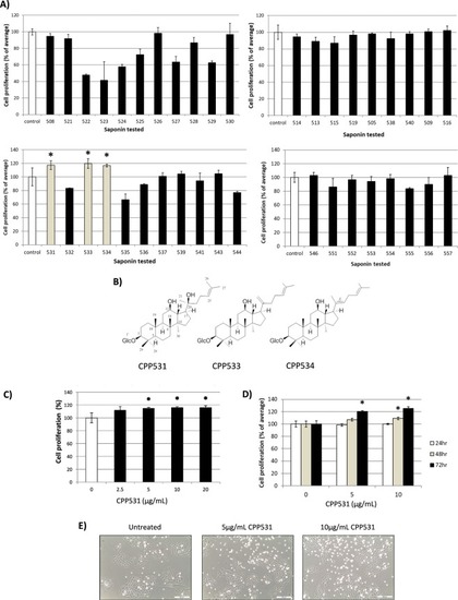
(A) Screening result of a compendium of 39 ginsenosides from P. ginseng for enhancers of myoblast proliferation. C2C12 myoblasts were treated with 5 µg/mL of each ginsenoside for 72 h. (B) Structure of the ′hit′ ginsenosides CPP531, ginsenoside Rk2 (designated as CPP533) and isoginsenoside Rh3 (designated as CPP534). (C) MTT assay showing the dose-dependent effect of CPP531 on C2C12 myoblast proliferation. Myoblasts were treated with CPP531 for 72 h. (D) MTT assay showing the time-dependent effect CPP531 treatment on C2C12 proliferation. (E) Micrographs of C2C12 myoblasts treated with CPP531 for 72 h. Scale bar = 200 µm. For (A–D): *p < 0.05 for significantly increased proliferation compared to vehicle treated myoblasts.

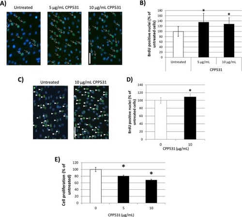
(A) BrdU immunostaining of C2C12 myoblasts treated with CPP531 for 72 h. Blue nuclei = DAPI, green nuclei = BrdU staining. Scale bar = 50 µm. (B) Quantification of BrdU labelled C2C12 nuclei. (C) BrdU immunostaining of human primary myoblasts treated with CPP531 for 72 h. Blue nuclei = DAPI, green nuclei = BrdU staining. Scale bar = 50 µm. (D) Quantification of BrdU labelled human myoblast nuclei. (E) Effect of 5 µg/mL CPP531 treatment for 72 h on proliferation in rat primary cardiac fibroblasts. For (B,D): *p < 0.05 for significantly increased BrdU nuclear staining compared to vehicle treated myoblasts; for (E) *p < 0.05 for significantly reduced proliferation compared to vehicle treated fibroblasts.

(A) Western blotting analysis of Akt expression in C2C12 myoblasts after treatment with 5 µg/mL CPP531 for 72 h. GAPDH was used as a loading control. (B) Quantification of Akt expression. (C) Western blotting analysis of phosphorylated Akt (P-Akt) expression in C2C12 myoblasts after treatment with 5 µg/mL CPP531 for 72 h. (D) Quantification of P-Akt expression. (E) RT-PCR analysis of p27Kip1 and p57Kip2 mRNA expression in C2C12 myoblasts after treatment with 5 µg/mL CPP531 or 5 μM BIO for 72 h. (F,G) Quantification of p27Kip1 and p57Kip2 mRNA expression. (H) Western blotting analysis of p27Kip1 and p57Kip2 expression in C2C12 myoblasts after treatment with 5 or 10 µg/mL CPP531, or 5 μM BIO, for 72 h. (I,J) Quantification of p27Kip and p57Kip2 protein expression. For (D): *p < 0.05 for significantly increased expression compared to vehicle treated myoblasts. For (F,G) and (I,J): *p < 0.05 for significantly decreased expression compared to vehicle treated myoblasts.

(A) MTT assay for cell proliferation in H9C2 cardiomyoblasts treated with 5 µg/mL CPP531 for 72 h. (B) Representative micrographs of the treated cardiomyoblasts. Scale bar = 200 μm. (C) BrdU labelling of rat neonatal cardiomyocyte nuclei after treatment with 5 µg/mL CPP531 5 µM BIO for 72 h. (D) RT-PCR analysis of p27Kip1 and p57Kip2 mRNA expression in rat neonatal cardiomyocytes after treatment with 5 µg/mL CPP531 or 5 μM BIO for 72 h. (E) Quantification of p27Kip expression. (F) Effect of 5 µg/mL CPP531 on cardiomyocyte proliferation in Tg(cmlc2:GFP) transgenic zebrafish. 20 hpf larvae were treated with CPP531 until 48 hpf and EdU staining was carried out at 72 hpf. Heart tissue and neighboring yolk sac are designated with dashed lines. White arrows designate double-labelled, proliferating cardiomyocytes. Two representative fish from the CPP531 treated and untreated groups are shown. For (A): *p < 0.05 for significantly increased proliferation compared to vehicle treated cardiomyoblasts. For (C): *p < 0.05 for significantly increased BrdU labelling compared to vehicle treated cardiomyocytes. For (C): *p < 0.05 for significantly decreased expression compared to vehicle treated cardiomyocytes.

(A) Representative echocardiograms from rats with sham MI (designated as ‘Normal’), MI and 7 d vehicle treatment (designated as ‘MI’) or MI and 7 d treatment with CPP531 (designated as ‘CPP531’). (B) Effect of CPP531 treatment on cardiac function related parameters. IVSTD: interventricular septal end diastole, LVIDd: left ventricular internal dimension, diastole, LVIDs: left ventricular internal dimension, systole, EF: ejection fraction, FS: fractional shortening and ESV: end systolic volume. n = 8 rats per treatment group; *p < 0.05 for significant improvement compared to vehicle treated rats with MI. (D) Masson’s trichrome staining of representative rat hearts. Red staining indicates cardiomyocytes and blue staining indicates fibrotic scar tissue. Scale bar = 1 mm. (E) Effect of CPP531 treatment on left ventricular wall thickness. *p < 0.05 for significantly greater ventricular wall thickness compared to vehicle treated rats with MI.

(A) Effect of CPP531 treatment on swim test motility time. *p < 0.05 for significant higher motility time compared to vehicle treated mice (designated as ‘no drug’). (B) Representative images of gastrocnemius muscle inflammation and fiber regeneration (indicated by the presence of small, centrally nucleated fibers) in vehicle treated mice and CPP531 treated mice 7 d after barium chloride injection. Scale bar = 100 µm. (C) Percentage of damaged gastrocnemius muscle tissue after 7 days. *p < 0.05 for significantly reduced damaged tissue compared to vehicle treated mice. (D) Muscle fiber diameter in vehicle treated mice and CPP531 treated mice after 7 d. *p < 0.05 for significantly larger diameter compared to vehicle treated mice. (E) Inflammatory cell count in the gastrocnemius muscle of untreated mice and CPP531 treated mice after 7 d. *p < 0.05 for significantly reduced inflammatory cell count compared to vehicle treated mice. (F) Representative images of the gastrocnemius muscle of untreated and CPP531 treated mice 14 d after barium chloride injection. Scale bar = 100 µm. (G) Percentage of damaged gastrocnemius muscle tissue after 14 d. *p < 0.05 for significantly reduced damaged tissue compared to vehicle treated mice. (H) Muscle fiber diameter in untreated mice and CPP531 treated mice at 14 d. *p < 0.05 for significantly increased diameter compared to vehicle treated mice. (I) Inflammatory cell count in the gastrocnemius muscle of untreated mice and CPP531 treated mice at 14 d. *p < 0.05 for significantly lower inflammatory cell count compared to the vehicle treated mice. n = 5 mice per treatment group.

Acknowledgments
This image is the copyrighted work of the attributed author or publisher, and ZFIN has permission only to display this image to its users. Additional permissions should be obtained from the applicable author or publisher of the image. Full text @ Sci. Rep.