- Title
-
Semaphorin3d mediates Cx43-dependent phenotypes during fin regeneration
- Authors
- Ton, Q.V., and Iovine, M.K.
- Source
- Full text @ Dev. Biol.
|
sema3d is differentially expressed in wild-type (top), alfdty86 (middle) and sofb123 (bottom). Left: whole mount in situ hybridization shows increased expression in alfdty86 and decreased expression in sofb123 compared to wild-type. Right: Cryosections reveal the tissue-specific localization of sema3d-expressing cells. Arrowheads point to skeletal precursor cells; arrows point to the basal layer of the epidermis (ble). EXPRESSION / LABELING:
|
|
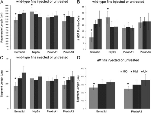
Morpholino-mediated gene knockdown of sema3d and its putative receptors. In all graphs, MO represents the particular gene-targeting morpholino; MM represents the particular 5 mis-match/control morpholino; UN represents uninjected/untreated fins. (A) Total regenerate length was measured. sema3d-knockdown and plxna3-knockdown cause reduced fin length (*); nrp2a-knockdown causes increased fin length (*), (B) the total number of H3P positive cells were counted. sema3d-knockdown causes reduced levels of cell proliferation (N); nrp2a-knockdown causes increased levels of cell proliferation (*), (C) segment length was measured in treated wild-type fins. sema3d-knockdown and plxna3-knockdown cause reduced segment length (*) and (D) segment length was measured in treated alfdty86 fins. sema3d-knockdown and plxna3-knockdown cause reduced segment length and rescue joint formation in alfdty86 (*). Statistically different data sets (*) were determined by the student′s t-test where p<0.05. By the student′s t-test, there is no statistical difference between MM and UN for any mismatch morpholino. Error bars represent the standard deviation. PHENOTYPE:
|
|
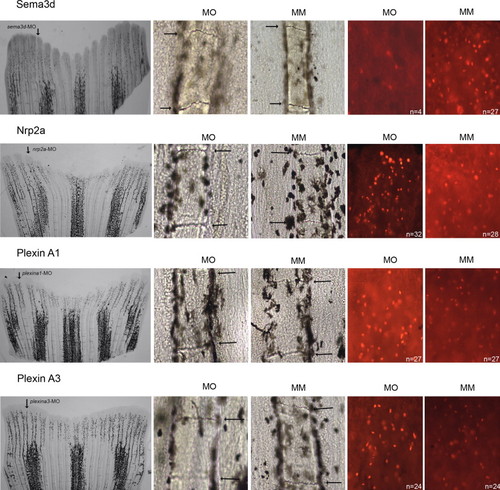
Representative images of morpholino-induced phenotypes. From left to right: representative whole fins following injection in the dorsal-most 5–6 fin rays (arrow); segment length in targeting morpholino-injected (MO) and in 5 mis-match morphlino-injected (MM); H3P-positive cells in targeting morpholino-injected (MO) and in 5 mis-match morphlino-injected (MM). PHENOTYPE:
|
|
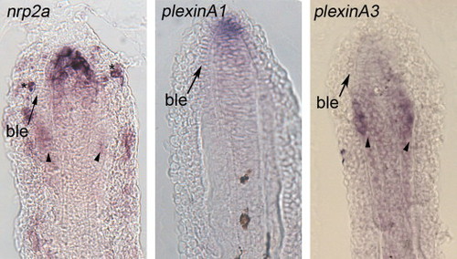
Gene expression of candidate receptors for Sema3d. Left: expression of nrp2a is primarily located in the distal blastema and in skeletal precursor cells. Staining of individual cells of the outer epithelial cells is also observed (N). Middle: expression of plxna1 is primarily in the distal blastema and in the distal cells of the basal epidermis. Right: expression of plxna3 is located in both the skeletal precursor cells and in the blastema. Arrows identify the basal layer of the epidermis (ble), arrowheads identify skeletal precursor cells. EXPRESSION / LABELING:
|
|
Nrp2a-knockdown effects are abrogated in sofb123. Nrp2a-mediated gene knockdown causes an increase in cell proliferation when Sema3d is present at typical levels. In sofb123, where sema3d expression is reduced, Nrp2a is unable to enhance the level of cell proliferation. MO, gene-targeting morpholino. MM, 5 mis-match/control morpholino. PHENOTYPE:
|

ZFIN is incorporating published figure images and captions as part of an ongoing project. Figures from some publications have not yet been curated, or are not available for display because of copyright restrictions. EXPRESSION / LABELING:
|
Reprinted from Developmental Biology, 366(2), Ton, Q.V., and Iovine, M.K., Semaphorin3d mediates Cx43-dependent phenotypes during fin regeneration, 195-203, Copyright (2012) with permission from Elsevier. Full text @ Dev. Biol.


