- Title
-
Regulatory interactions specifying Kolmer-Agduhr interneurons
- Authors
- Yang, L., Rastegar, S., and Strähle, U.
- Source
- Full text @ Development
|
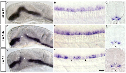
nkx2.2a, nkx2.2b and nkx2.9 are expressed in overlapping domains. (A-I) Head (A,D,G) and trunk (B,E,H lateral view; C,F,I transverse section) of a 24 hpf zebrafish embryo hybridized to nkx2.2a (A-C), nkx2.2b (D-F) and nkx2.9 (G-I) antisense probes. Whereas expression of nkx2.2a (A) along the ventral brain is continuous, the expression domains of nkx2.2b and nkx.2.9 show a gap (D,G, asterisk) in the prethalamic and thalamic areas adjacent to the zona limitans intrathalamica. All three genes are expressed in the lateral floor plate of the trunk. In the spinal cord (B,E,H), the expression of nkx2.2a and nkx2.9 is discontinuous, with gaps of non-expressing cells (B,H, black arrowheads), whereas all cells of the lateral floor plate appear to express nkx2.2b (E). All three genes are restricted to the lateral floor plate (C,F,I). Orientation of whole-mount embryos (A,B,D,E,G,H): anterior left, dorsal up. Lateral views of the head (A,D,G) and the spinal cord are at the level of the hindgut extension. Transverse sections (C,F,I) are at the level of the hindgut extension, dorsal up. m, medial floor plate; n, notochord. Scale bar: 100 μm in A,D,G; 25 μm in B,C,E,F,H,I. EXPRESSION / LABELING:
|
|
tal2 expression in the lateral floor plate depends on nkx2.2a, nkx2.2b and nkx2.9. (A,B) Control (A) and Mo-nkx2.9-injected (B) zebrafish embryo. Knockdown of nkx2.9 leads to a reduction of tal2-positive cells in the lateral floor plate (arrow) but dorsal tal2-positive cells are unaffected (arrowhead). (C,D) Control (C) and Mo-nkx2.9/Mo-nkx2.2a-injected (D) embryos. There is a reduction of tal2-positive cells in the lateral floor plate, whereas tal2-expressing cells (arrowhead) further dorsal in the spinal cord are unaffected in the double-knockdown embryos. (E,F) Control (E) and Mo-nkx2.9/Mo-nkx2.2b-injected (F) embryos. There is a reduction of tal2-positive cells in the lateral floor plate (arrow) in the double-knockdown embryos. (G,H) Control (G) and Mo-nkx2.2a/Mo-nkx2.2b-injected (H) embryos. Double knockdown of nkx2.2a and nkx2.2b caused little reduction of tal2-positive cells (arrow) in the lateral floor plate. (I,J) Control (I) and triple-knockdown (J) embryos. Knockdown of all three Nkx2 genes abolished tal2-positive cells in the lateral floor plate (arrow). tal2-expressing cells (arrowhead) further dorsal in the spinal cord were unaffected in these knockdown embryos. (K) Cross-section through the trunk at hindgut extension of a triple-knockdown embryo. tal2-expressing cells in the lateral floor plate (arrow) are missing, whereas more dorsally located tal2-positive cells are present (arrowhead). Note that controls represent injections of mismatch morpholinos or combinations thereof (Table 1). Representative lateral views of the spinal cord over the hindgut extension are shown. Embryos (24 hpf) are shown with anterior left, dorsal up. Scale bar: 25 μm. |
|
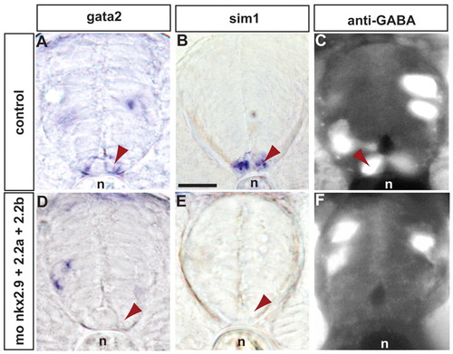
gata2-expressing, sim1-expressing and GABAergic cells are missing in the lateral floor plate of triple-knockdown zebrafish embryos. (A-F) Transverse sections through control morpholino-injected (A-C) and triple-injected (Mo-nkx2.9, Mo-nkx2.2a, Mo-nkx2.2b) (D-F) embryos were hybridized to gata2 (A,D) or sim1 (B,E) antisense probe or anti-GABA antibody (C,F). Cells in the lateral floor plate (arrowhead) that express gata2 and sim1 or that are GABAergic are abolished by triple knockdown of nkx2.2a, nkx2.2b and nkx2.9 (D-F), whereas cells expressing the same markers more dorsally appear unaffected. Embryos were 24 (A,C,D,F) or 48 (B,E) hpf. All transverse sections were cut at the level of the yolk extension. n, notochord. Scale bar: 25 μm. |
|
Mapping co-expression of marker genes and GABA in the zebrafish lateral floor plate. (A-C) nkx2.9 (A) and tal2 (B) mRNA expression and a merged view (C). nkx2.9-positive cells (arrowheads) co-express tal2. (D-F) GABA immunohistochemistry (D), tal2 in situ hybridization (E) and a merged view (F). tal2-expressing cells (arrowheads) are GABAergic. (G-I) gata2 (G) and tal2 (H) mRNA expression and a merged view (I). gata2 and tal2 mRNAs (arrowheads) are co-expressed in the lateral floor plate. More dorsally in the spinal cord, not all gata2-positive cells are tal2-positive. (J-L) gata3 (J) and tal2 (K) mRNA expression and merge (L). gata3 mRNA-expressing cells co-express tal2 mRNA in the lateral floor plate. However, more dorsally, only a proportion of gata3-positive cells also expresses tal2 mRNA. The tal2-negative, gata2- and gata3-positive cells are probably V2b/VeLD interneurons (Batista et al., 2008). (M-O) sim1 (M) and tal2 (N) mRNA expression and merge (O). sim1-expressing interneurons and tal2-expressing cells in the lateral floor plate are distinct in most cases. Only in 25% of cells did we find co-expression of the two markers. (A-F) Projections of several sections. (G-O) Single confocal planes. Embryos were 24 (A-L) or 36 (M-O) hpf. Dorsal up, anterior left. Scale bar: 50 μm. |
|
gad67 expression in KA′ and KA′ cells is differentially dependent on tal2 and olig2. (A,B) Zebrafish embryos injected with a mixture of mismatch morpholinos (A) or a cocktail of morpholinos directed against nkx2.2a, nkx2.2b and nkx2.9 mRNA (B). The nkx2 cocktail abolished gad67 expression in KA′ cells (arrow) in the lateral floor plate (B). gad67 expression in KA′ and VeLD cells (arrowhead) was not affected by the knockdown of the Nkx2 genes. (C,D) Embryos injected with a mismatch morpholino (C) or with a morpholino directed against tal2 mRNA (D). gad67 expression in KA′ cells (arrow) was abolished in the tal2 morphants, whereas gad67 expression in KA′ and VeLD cells (arrowhead) appeared to be normal. (E,F) Embryos injected with a mismatch morpholino (E) or a morpholino directed against olig2 mRNA (F). The strong gad67 expression in dorsally located cells (arrowhead) was abolished by knockdown of olig2. Expression in KA′ cells (arrow) and the low-level expression in what are presumably V2 interneurons was not affected by knockdown of olig2 expression. (G,H) Embryos injected with either a mix of tal2 and olig2 mismatch control morpholino (G) or with morpholinos directed against tal2 and olig2 mRNA (H). Co-injection of the tal2 and olig2 morpholinos abolished gad67 expression in both the KA′ (arrowhead) and KA′ (arrow) interneurons. Expression of gad67 in some dorsally located cells, which probably represent V2 interneurons expressing gad67 at low levels, still persisted in the spinal cord of double-injected morphants. Embryos were 24 hpf. Anterior left, dorsal up. Scale bar: 25 μm. |
|
gata2 and gata3 expression in the lateral floor plate requires Nkx2 genes but not tal2. (A,B) Zebrafish embryos injected with a mix of mismatch morpholinos (A) or morpholinos directed against nkx2.2a, nkx2.2b and nkx2.9 mRNA (B). gata3-expressing cells are not detectable in the lateral floor plate (arrow) in triple morphants, whereas dorsally located gata3-expressing cells (arrowhead) are present. (C,D) Embryos injected with a mismatch control morpholino (C) or a morpholino directed against tal2 mRNA (D). tal2 knockdown does not affect gata3 expression, indicating that tal2 does not regulate gata3. (E,F) Embryos were injected with a mismatch morpholino (E) or a morpholino directed against tal2 mRNA (F). gata2 expression is not abolished by knockdown of tal2 expression. Embryos were 24 hpf. Anterior left, dorsal up. Scale bar: 25 μm. |
|
gata2 and gata3 differentially regulate gad67 expression in KA′ and KA′ interneurons. (A-D) Zebrafish embryos injected with mismatch morpholino (A,C) or with morpholinos directed against gata2 mRNA (B,D). gata2 abolishes tal2 expression (B) as well as gad67 expression (D) in the lateral floor plate (arrow). Thus, correct differentiation of KA′ cells in the lateral floor plate requires gata2 function. (E-H) Embryos injected with mismatch morpholino (E,G) or morpholinos directed against gata3 mRNA (F,H). tal2 (E,F) and gad67 (G,H) expression in KA′ cells is abolished by gata3 knockdown (arrowhead), whereas expression in KA′ cells is unaffected (arrow). Embryos were 24 hpf. Anterior left, dorsal up. Scale bar: 25 μm. |
|
Lateral floor plate identity depends on nkx2.2a, nkx2.2b and nkx2.9. (A,B) Control (A) and Mo-nkx2.2a/Mo-nkx2.2b/Mo-nkx2.9-injected embryo (B) stained with gata2 antisense probe. Triple knockdown abolished gata2-positive cells in the lateral floor plate (arrow) completely. gata2-expressing cells (arrowhead) located dorsally in the spinal cord were unaffected. (C,D) Control (C) and nkx2 triple-knockdown (D) embryos hybridized to sim1 antisense probe. Knockdown of all three Nkx2 genes deleted sim1 expression in the lateral floor plate (arrow). (E,F) Control (E) and triple-knockdown (F) embryos labeled with anti-GABA antibody. Anti-GABA staining was completely lost in the lateral floor plate (arrow) in the triple-knockdown embryo. GABAergic neurons (arrowhead), which are located more dorsally, are not affected by knockdown of the three Nkx2 genes. (G,H) Control (G) and triple-knockdown (H) embryos stained with pax6.1 antisense probe. The expression of pax6.1 is not affected by the triple knockdown. (I,J) Control (I) and triple-knockdown (J) embryos stained with pax6.2 probe. As in the case of pax6.1, pax6.2 is not expanded into the lateral floor plate in Nkx2 triple-knockdown embryos. (K,L) Control (K) and triple-knockdown (L) embryo hybridized to olig2 probe. olig2-positive cells are found ectopically in the lateral floor plate in nkx2 triple-knockdown embryos. The dashed line indicates the border between the lateral floor plate and more dorsally located cells. Arrowheads outline the increased width of the olig2 expression domain in the morphants. pax6.1 and pax6.2 expression does not expand into the lateral floor plate in triple-knockdown embryos, whereas olig2-expressing cells were aberrantly found in the lateral floor plate in these knockdown embryos. Representative lateral views of the spinal cord over the hindgut extension are shown. Embryos are 48 hpf (C,D) and 24 hpf (A,B,E-L) and are oriented with anterior left and dorsal up. Scale bar: 25 μm. |
|
The differentiation of V3 interneurons depends on the knockdown of nkx2.9, nkx2.2a and nkx2.2b but not on gata2. (A,B) Control (A) and Mo-nkx2.2a/Mo-nkx2.2b-injected (B) embryos stained with sim1 probe. In contrast to the triple knockdown including nkx2.9 (see Fig. 5B,E, Fig. S1C,D), the double knockdown does not affect sim1 expression (arrow) indicating that nkx2.9 is also crucial for the development of V3 interneurons. (C,D) Control (C) and Mo-gata2-injected (D) embryos hybridized to the vglut2.1 probe. As with sim1 expression, the expression of vglut2.1 (arrow) is not affected by knockdown of gata2 indicating that V3 interneurons are independent of gata2 function. (E,F) Control (E) and Mo-nkx2.2a, Mo-nkx2.2b, Mo-nkx2.9 injected (F) embryos stained with vglut2.1 antisense probe. The vglut2.1-expressing cells (arrow) located in the lateral floor plate are missing in the knockdown embryo, whereas more dorsally located glutamatergic cells are unaffected. (G,H) Forty-eight-hour-old control (G) and Mo-gata2-injected (H) embryos stained with tal2 probe. The expression of tal2 (arrow) in the lateral floor plate is abolished completely, whereas more dorsally located tal2-expressing cells (arrowhead) are unaffected. (I,J) Forty-eight-hour-old control (I) and Mo-olig2/Mo-tal2 double-injected (J) embryos hybridized to gad67 probe. gad67-expressing cells (arrow) in the lateral floor plate and more dorsal regions (arrowhead) are missing in knockdown embryos. This shows that these morpholinos are also highly effective at 48 hpf. (K,L) Control (K) and Mo-olig2-injected (L) embryos hybridized to gata3 antisense probe. gata3-expressing KA′ cells (arrowhead) are missing in knockdown embryos. (M,N) Control (M) and Mo-gata2-injected (N) embryos stained with gata3 antisense probe. gata3 expression in the lateral floor plate (arrow) but not in the KA′ cells (arrowhead) are abolished in morphants. Embryos are 48 hpf (A-J) or 24 hpf (K-N) and oriented anterior left and dorsal up. Scale bar: 25 μm. |
|
Expression of vesicular glutamate transporter 2.1 (vglut2.1) in sim1-positive cells. (A) sim1 expression; (B) vglut2.1 expression; (C) merged view of A and B. Most sim1-positive cells co-express vglut2.1, indicating that they are indeed similar to mouse V3 interneurons. All embryos are 36 hpf and oriented anterior left and dorsal up. Scale bar: 25 μm. |


