- Title
-
Neuregulin-mediated ErbB3 signaling is required for formation of zebrafish dorsal root ganglion neurons
- Authors
- Honjo, Y., Kniss, J., and Eisen, J.S.
- Source
- Full text @ Development
|
erbb3b and erbb2 mutants lack DRG and sympathetic neurons. (A-C) Elavl antibody labeling reveals DRG (arrowheads) and sympathetic neurons (asterisks). DRGs are segmentally arranged, whereas at this stage, sympathetics are not segmental. DRG and sympathetic neurons are present in wild-type embryos (A) but are absent from erbb3b (B) and erbb2 (C) mutants at 7 dpf. (D-F) Elavl-positive enteric neurons appear normal in erbb3b (E) and erbb2 (F) mutants compared with wild-type embryos (D). (G-I) TH antibody labeling reveals sympathetic neurons. Whole-mount labeling of 8 dpf larvae showing that sympathetic neurons (asterisks) are present in wild type (G) but absent from erbb3b (H) and erbb2 (I) mutants. (J-L) Whole-mount labeling of 31 hpf embryos with a neurog1 riboprobe reveals nascent DRG neurons. Nascent DRG neurons (arrowheads) are segmental in wild-type embryos (J) but absent from erbb3b (K) and erbb2 (L) mutants. (M-O) A neurod riboprobe reveals DRG neurons at 36 hpf. DRG neurons (arrowheads) are segmental in wild-type embryos (M) but absent from erbb3b (N) and erbb2 (O) mutants. Lateral views; anterior towards the left. Scale bar: 20 μm. |
|
Neural crest cell migration is initially normal but later disrupted in erbb3b mutants. (A-H) Whole-mount labeling using crestin (A,B,E-H) or sox10 (C,D) riboprobes of wild-type embryos (A,C,E,G) and erbb3b mutants (B,D,F,H). At 24 hpf, both probes show that NC cell migration appears normal in erbb3b mutants (B,D) and in wild type (A,C). At 27 (E,F) and 30 (G,H) hpf, NC cells form segmental streams ventral of the notochord in wild type (E,G). By contrast, in erbb3b mutants (F,H) there are no streams at these stages, and NC cell migration is disrupted. Broken lines represent dorsal aspect of the notochord. Lateral views, anterior towards the left. Scale bar: 20 μm. EXPRESSION / LABELING:
PHENOTYPE:
|
|
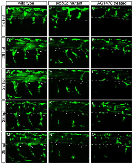
In erbb3b mutants, neural crest cells fail to pause in the location where DRGs normally form. Single frames taken from confocal time-lapse movies of 22-70 hpf Tg(sox10:GFP) zebrafish. In wild-type embryos (A,D,G,J,M), NC cells pause at the dorsal and ventral aspects of the notochord for more than 24 hours. By contrast, in erbb3b mutants (B,E,H,K,N) and in AG1478-treated wild type (C,F,I,L,O), NC cells did not pause and abnormally migrated away. Arrowheads indicate pausing NC cells; asterisks indicate places where pausing NC cells are missing. Broken lines indicate dorsal aspect of notochord. Scale bar: 20 μm. |
|
ErbB signaling is required during neural crest migration. Control embryos and embryos treated with AG1478 for various time periods were labeled with Elavl antibody at 4 dpf to examine DRG formation. (A-F) Somites 4-18 of inhibitor-treated (A,C,E) and control (B,D,F) embryos. DRG neurons were missing between somites 5 and 11 when embryos were treated from 8-14 hpf (A) and DRG neurons were missing throughout the body when embryos were treated from 18-30 hpf (C). DRG neurons were present anteriorly but missing posterior of somite 13 when embryos were treated from 24-30 hpf (E). DRG neurons were present in all segments in control embryos. Arrowheads in A, C and E indicate DRG neurons. (G,H) Graphs showing average percentage of fish with DRG neurons at each of three axial levels when embryos were treated for various time periods. In control embryos, DRG neurons were present in 90-100% of fish for all experiments. At least 30 embryos were counted in each experiment. Scale bar: 50 μm. EXPRESSION / LABELING:
|
|
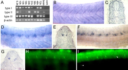
nrg1 alone is not required for DRG formation. (A) RT-PCR showing expression of each nrg1 isoform at various developmental stages and in the adult brain. (B,D,F) Expression patterns of each isoform at 24 hpf shown in whole-mount. (C,E,G) Cross section of expression pattern of each isoform respectively. (B,C) Type I is expressed weakly in somites. (D,E) Type II and (F,G) type III are expressed strongly in ventral spinal cord neurons. (E,F) Elavl labeling at 2 dpf. (H,I) Control MO-injected embryos (H) had segmental DRG neurons. nrg1 MO-injected embryos (I) had only a slight decreased in DRG neuron number; arrowhead indicates absent DRG, asterisk shows mislocalized DRG. Scale bar: 40 μm. EXPRESSION / LABELING:
PHENOTYPE:
|
|
Zebrafish has two nrg2 genes. (A) RT-PCR showing expression of nrg2a and nrg2b at various developmental stages and in the adult brain. Whole-mount labeling of wild-type embryos showing nrg2a expression in somites (B, cross section in C) and nrg2b expression in spinal neurons at 24 hpf (D, cross section in E). Scale bar: 20 μm. EXPRESSION / LABELING:
|
|
nrg2a alone is unnecessary for DRG neuron formation. DRG neurons labeled with Elavl antibody at 4 dpf. DRG neurons in controls (A,C). DRG neurons are missing from some segments of nrg2a MO-injected embryos (B) but appear normal in nrg2b MO-injected embryos (D). Asterisks indicate absent DRG neurons in B. (E) RT-PCR showing that nrg2a MOs cause incorrect mRNA splicing through 3 dpf, but by 4 dpf, both incorrectly-spliced and correctly-spliced transcripts are present. (F) RT-PCR showing that both the nrg2a and nrg2b MOs cause incorrect splicing at 25 hpf. Scale bar: 20 μm. |
|
nrg1 and nrg2a act redundantly during DRG neuron formation. (A,B) Elav1 antibody labeling at 4 dpf. Co-injection of nrg1 and nrg2a MOs resulted in absence of DRG neurons (B), whereas DRG neurons are normal in controls (A). (C-H) Whole-mount wild-type (C,E,G) and nrg1 plus nrg2a MO-injected embryos (D,F,H) labeled with crestin riboprobe at 24 hpf (C,D), 27 hpf (E,F) and 30 hpf (G,H). Like erbb3b and erbb2 mutants, double MO-injected embryos have normal early NC migration (C,D) but, at later stages, NC migration is disrupted (E-H). Scale bar: 20 μm. |


