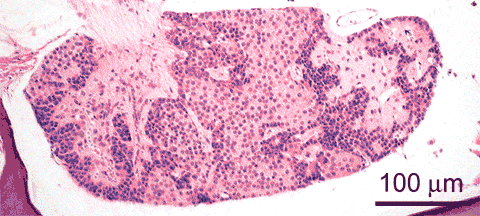
Adult male zebrafish; H&E staining sagittal view
The pituitary is composed of nervous tissue, bundled in the neurohypophysis
 , and of endocrine cells, clustered in the adenohypophysis
 . The neurohypophysis connects the brain through the stalk of the hypophysis
 with the adenohypophysis, in which it branches off.
 Classically, the adenohypophysis is subdivided in the rostral pars distalis (also known as the pro-adenohypophysis), the proximal pars distalis (meso-adenohypophysis), and the pars intermedia (meta-adenohypophysis); see Hibiya 1982, for a diagrammatical comparison of these substructures in several fish species 1.
These parts contain specialized cell types, which, in the zebrafish, cannot be distinguished on routine sections.
On sagittal H&E stained sections of the pituitary in the zebrafish, the main substructures which can be distinguished in the adenohypophysis are the basophilic small cells which line the neurohypophysis , and the large cells which constitue the main body of the adenohypophysis
 , of which acidophilic cells
 stand out. An example of distinct morphology of a part of the pituitary is shown on the PAS stained sections of guppy after β-HCH.
See below for further discussion of various cell types of the adenohypophysis.
On sagittal H&E stained sections of the pituitary in the zebrafish, the main substructures which can be distinguished in the adenohypophysis are the basophilic small cells which line the neurohypophysis
, and the large cells which constitue the main body of the adenohypophysis, of which acidophilic cells stand out. An example of distinct morphology of a part of the pituitary is shown on the PAS stained sections of guppy after β-HCH.See below for further discussion of various cell types of the adenohypophysis.
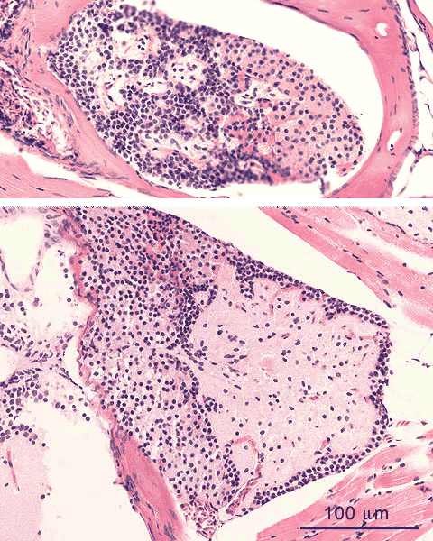
Adult male zebrafish; H&E staining coronal view
On coronal H&E stained sections of the pituitary in the zebrafish, the main substructures which can be distinguished in the adenohypophysis are the basophilic small cells
 which line the neurohypophysis
 , and the large cells
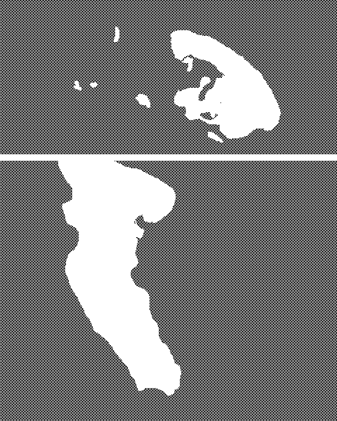 which constitue the main body of the adenohypophysis
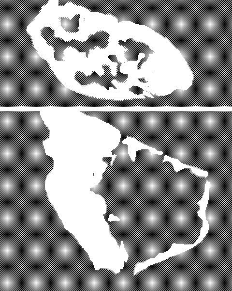 , of which acidophilic cells
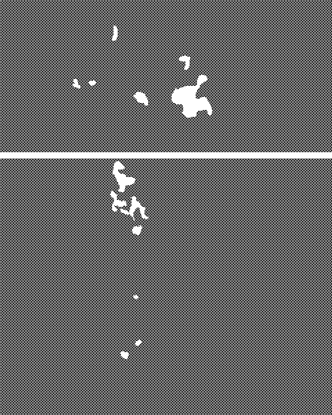 stand out. An example of distinct morphology of a part of the pituitary is shown on the PAS stained sections of guppy after β-HCH.
See below for further discussion of various cell types of the adenohypophysis.
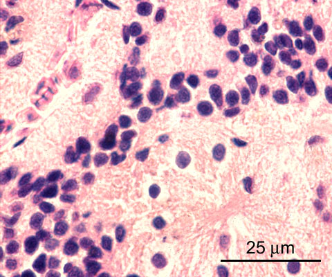
Adult male zebrafish; H&E staining details - neurohypophysis
This image shows the intersection between neurohypophysis and adenohypophysis.
The neurohypophysis
 consists of nerve fibers
 and scattered pituicytes
 , which are glia cells.
The basophil cells
 which line the neurohypophysis in 3-4 layers are ACTH-cells (corticotrops)1.
A capillary with erythrocytes
 crosses in the upper left corner.
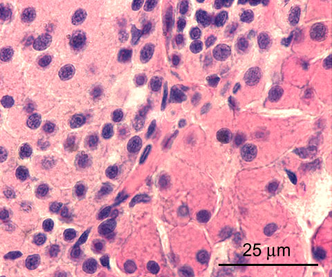
Adult male zebrafish; H&E staining details - adenohypophysis
The adenohypophysis contains several cell types, which have differential histochemical stainability patterns. With H&E, as applied in these sections, acidophilic, basophilic, and chromophobe cells are distinghuised. Acidophilic cells
 are large and show in clusters, as do chromophobe cells
 . In zebrafish, basophilic cells are small, and show either in strands spanning 3-4 cell layers
 , lining the neurohypophysis, or scattered in the adenohypophysis
 . Other species may show a different organisation of these cell types (see Hibiya 19821). Hibiya also refers to histochemical stainings to identify various cell types, according to their specific expression patterns. In this way, prolactin producing cells may be differentiated from cells producing growth hormone (somatotropic hormone), thyrotrop hormone (TSH), gonadotrop hormones, melanotropin (MSH), and adrenocorticotrop hormone (ACTH). However, immunohistochemical staining techniques are now available for the idenfication of hormone production (see Rodriguez-Gomez 2001 for detailed mapping in tunafish2).
A capillary with associated stromal tissue
 crosses the section.
- Hibiya-T (ed.). An Atlas of Fish Histology - Normal and Pathological Features. Kodansha, Tokyo / Gustav Fisher Verlag, Stuttgart - New York; 1982.
- Rodriguez-Gomez-FJ, Rendon-Unceta-MC, Pinuela-C, Munoz-Cueto-JA, Jimenez-Tenorio-N, Sarasquete-C. Immunocytohistochemical characterization of pituitary cells of the bluefin tuna, Thunnus thynnus L. Histol.Histopathol.16:443-451;2001.