
- Title
-
Rifamycin O, An Alternative Anti-Mycobacterium abscessus Agent
- Authors
- Hanh, B.T.B., Park, J.W., Kim, T.H., Kim, J.S., Yang, C.S., Jang, K., Cui, J., Oh, D.C., Jang, J.
- Source
- Full text @ Molecules
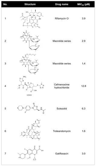
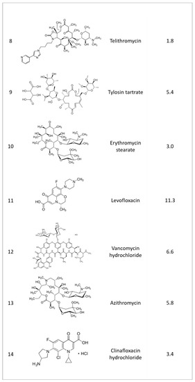
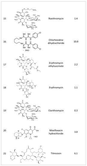
Structure and 50% minimum inhibitory concentration (MIC50) values of the 23 most potent |
Structure and 50% minimum inhibitory concentration (MIC50) values of the 23 most potent |
Structure and 50% minimum inhibitory concentration (MIC50) values of the 23 most potent |

Structure and 50% minimum inhibitory concentration (MIC50) values of the 23 most potent |
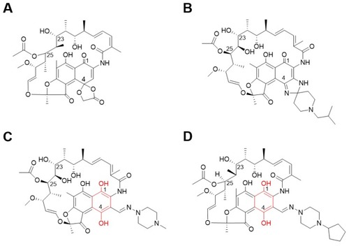
Chemical structures of rifamycin analogs. Rifamycin O ( |
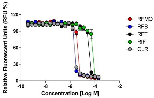
In vitro activity of rifamycin O. The activity of rifamycin O (RFM O) against |
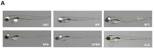
Evaluation of in vivo RFM O activity on |
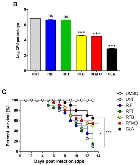
Evaluation of in vivo RFM O activity on PHENOTYPE:
|
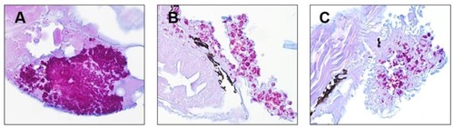
Multiple acid-fast bacilli are present in the ZF. ZF with |