
- Title
-
not really finished is crucial for development of the zebrafish outer retina and encodes a transcription factor highly homologous to human Nuclear Respiratory Factor-1 and avian Initiation Binding Repressor
- Authors
- Becker, T.S., Burgess, S.M., Amsterdam, A.H., Allende, M.L., and Hopkins, N.
- Source
- Full text @ Development
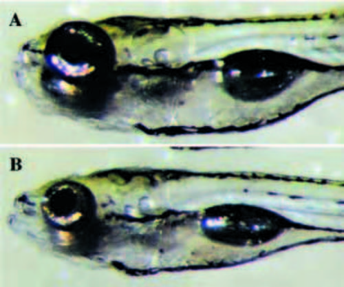
Micrographs of live zebrafish larvae at 120 hpf. (A) Wild-type individual, (B) nrf/nrf mutant. Note the much smaller eyes in the mutant. PHENOTYPE:
|
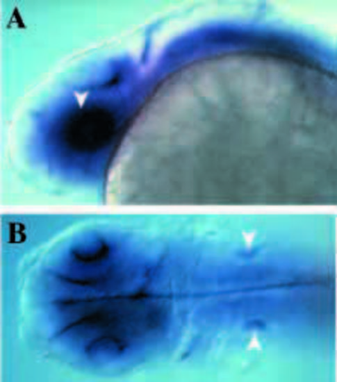
In situ hybridization illustrating the expression of nrf in the 24h embryo. (A) Lateral view: expression is very strong in the developing eye (arrowhead) and the CNS, including the spinal cord. (B) Dorsal view: expression is strongest in the eyes and brain, as well as in the ears (arrowheads). EXPRESSION / LABELING:
|
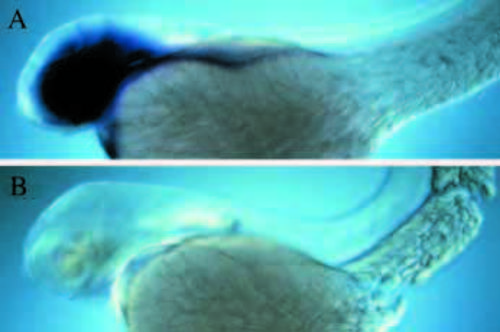
Whole-mount in situ hybridization with the Nrf antisense probe at 48 hpf. (A) Wild-type, note strong expression in the eyes and the brain. (B) nrf/nrf homozygous sibling. nrf mRNA cannot be detected. |
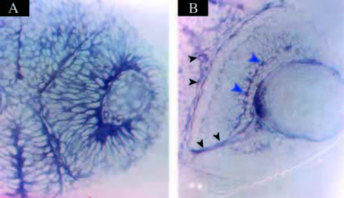
Sections of in situ hybridizations using the Nrf probe in 24 hour (A) and 48 hour (B) retinae. Note that at 24 hpf, expression of nrf is detected in every cell, whereas at 48 hpf, expression is highest in the ganglion cell layer (large blue arrowheads), optic nerve and optic tracts (small black arrowheads), while very low in the PR layer. |

Cross sections through retinae at 50 hpf prepared from whole mounts stained with monoclonal antibody Zn5. (A) Wild type; note the Zn5-positive retinal ganglion cells and the optic nerve (red arrowhead). In the back of the retina, a layer of prospective photoreceptor cells can be distinguished (yellow arrowheads). (B) nrf/nrf sibling, note that Zn5-positive cells and optic nerve are present (red arrowhead), but that layering of the retina is not as apparent. |

Frontal sections through retinae at 72 hpf stained with mAB Fret43, which stains photoreceptors after their final mitosis. (A) Wild-type retina is already layered into the major three layers and photoreceptors begin appearing in the ventral patch (red arrowheads). (B) nrf/nrf mutant also shows differentiating red/green cones (red arrowheads) but, in addition in the mutant, multiple rounded cells, not integrated into the tissue are distinguished (small black arrowheads). EXPRESSION / LABELING:
PHENOTYPE:
|
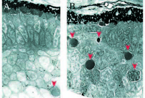
Electron micrographs of frontal sections from retinae at 76 hpf. (A) Wild-type retina; retinal pigmented epithelium is to the top. Note the already formed PR cell layer with outer segments. (A) Single apoptotic cell can be seen (arrowhead). (B) nrf/nrf sibling, note the discontinuous PR layer and high numbers of apoptotic profiles (arrowheads). PHENOTYPE:
|

Whole-mount TUNEL stains of 80 hpf retinae. (A) In wild type, a few cells undergoing cell death (arrowheads) are detected. (B) In the mutant, large clusters of apoptotic retinal cells are evident. PHENOTYPE:
|

In situ hybridization using four different opsin probes comparing wild type and mutant at 100 hpf. (A,B) Red opsin, expression is detected in the mutant (B). (C,D) Blue opsin; (C) wild type, (D) mutant: expressing cells are found (arrowheads). (E,F) Goldfish UV opsin; (E) weak expression is seen in the wild type but (F) not in the mutant. (G,H) Rod opsin; (G) in the wild type, rod opsin expression at this stage is seen mostly in the ventral retina and (H) is detected in the mutant (arrowhead). EXPRESSION / LABELING:
|

Frontal plastic sections through wild-type (A) and mutant (B) retinae at 120 hpf. Note the almost complete absence of photoreceptors in the mutant at this stage, as well as deficiencies in the inner nuclear layer, particularly in the peripheral retina. However, small clusters of photoreceptors (arrowheads) are discernible. PHENOTYPE:
|

Electron micrographs of frontal sections from larvae at 120 hpf. (A) Wild type; retinal pigmented epithelium is seen at the top left, directly adjacent to the outer segments of the cones (arrowheads) as well as of rods (interstitial cells). (B) In the mutant, small clusters of differentiated photoreceptors can be seen, complete with (deformed) outer segments (arrowheads). PHENOTYPE:
|