- Title
-
Notochord regulates cardiac lineage in zebrafish embryos
- Authors
- Goldstein, A.M. and Fishman, M.C.
- Source
- Full text @ Dev. Biol.
|
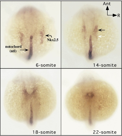
Relationship of Nkx2.5 expression and the anterior notochord. Whole mount in situ hybridization was performed on embryos fixed at the stages shown, and labeled with antisense probes for Nkx2.5 and ntl. Bilateral expression of Nkx2.5 is evident at the 6-somite stage. At the 14-somite stage the Nkx2.5 domain is more medially positioned (black arrow), and expression in its posterior extent (white arrow), that portion lateral to anterior notochord, is diminishing. At 18-somite stage, the caudal ends of the bilateral tubes begin to fuse just anterior to the notochord tip. Fusion is complete by the 22-somite stage, appearing as a ring anterior to the notochord. |
|
Laser ablation of anterior notochord. Dorsal view of a wild-type embryo at the 12-somite stage showing a normal anterior notochord before (A) and 10 min after (B) laser ablation (arrow denotes tip of notochord, black line marks first somite for reference). C and D demonstrate histologically the notochord (nt) before and after ablation, respectively. |
|
Posterior expansion of Nkx2.5 expression follows anterior notochord ablation. (A) Wild-type embryo at the 20-somite stage demonstrating normal pattern of Nkx2.5 expression (region of interest is magnified in the right panel). (B) Nkx2.5 expression in a 20-somite stage embryo whose anterior notochord had been ablated at the 8-somite stage, demonstrating posterior expansion of Nkx2.5 expression (white arrow; magnified view shown in right panel). |
|
Nkx2.5 expression in ntl mutant embryos. Nkx2.5 expression is shown in wild-type (wt) embryos at the 17-, 18-, and 21-somite stages. pax-b expression at the midbrain–hindbrain junction anteriorly and the otic vesicle posteriorly serves as a marker of anterior–posterior distance for comparison. Marked posterior extension of Nkx2.5 gene expression is evident in ntl embryos at the 21-somite stage (lower-right panel) when compared to normal embryos (lower-left panel). |
|
Notochord ablation redirects cardiac cell fate. The left panel schematics show the site of dye activation relative to the Nkx2.5-expressing zone and the notochord in a 14-somite stage embryo. The middle panels show the location of activated dye as fluorescence images (the white arrow denotes tip of notochord). The right panels show the heart region at prim-5 stage. (A) Normally, cells anterior to the notochord generate progeny in the heart (right panel). (B) Lateral mesoderm cells more posteriorly, next to the notochord, do not normally contribute progeny to the heart. (C) After notochord ablation, cells in the same posterior region as in B now contribute progeny to the heart. |
Reprinted from Developmental Biology, 201, Goldstein, A.M. and Fishman, M.C., Notochord regulates cardiac lineage in zebrafish embryos, 247-252, Copyright (1998) with permission from Elsevier. Full text @ Dev. Biol.


