
- Title
-
A series of no isthmus (noi) alleles of the zebrafish pax2.1 gene reveals multiple signaling events in development of the midbrain-hindbrain boundary
- Authors
- Lun, K. and Brand, M.
- Source
- Full text @ Development
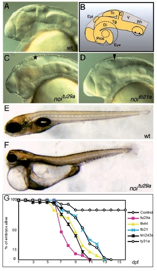
Lateral views of wild-type and noi mutant embryos. (A) Wildtype embryo at 24 hours pf. (B) Schematic drawing of the structures seen in A. (C) Strong noitu29a mutant, which lacks MHB, tectum (asterisk) and cerebellum. (D) Weak noitb21 phenotype; a partially formed tectum is observed, the caudal end marked by the arrowhead. (E) A wild-type embryo at day 7. A homozygous mutant for noitu29a of the same age is seen in (F), showing severe oedema of the pericard and gut epithelium, causing delayed development. (G) Survival rates of different noi alleles. n=30 mutant embryos per allele were analyzed. The difference between the strong noitu29a allele and the weaker alleles is clearly visible. The mutants never feed and die within 2 weeks. c, cerebellum, di, diencephalon, epi, epiphysis, pros, prosencephalon, rh, rhombencephalon, tc, tectum, tg, tegmentum, v, ventricle. |
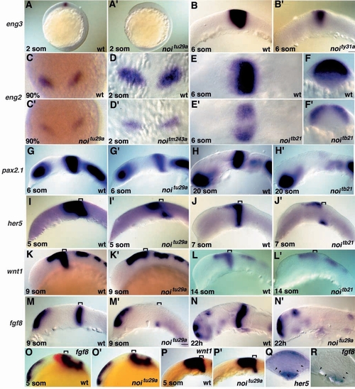
Requirement for Pax2.1 in early MHB development. Wholemount ISH; embryos shown in one row are stained with the marker depicted on the left side. Stages and genotypes are noted; shown are lateral views, anterior to the left, except where indicated. A prime (′) indicates mutants. (A) eng3 expression is not activated in noitu29a. (B) In weak noity31a mutants expression fades away from ventral to dorsal. (C,D) Weak transient eng2 expression in the MHB primordia, dorsal views, anterior to the top; in noitu29a (C) the expression is very faint and lost at the end of epiboly; in strong noitm243a mutants (D) expression initiates normally, fades away around 2 somites, and is lost later in development. (E) Dorsal views and (F) optical cross sections through the eng2 domain of wild-type embryo and weak noitb21 mutants. Expression persists dorsally. (G,H) Expression of pax2.1 as a marker for MHB development is initiated normally in noitu29a mutants, and is gradually eliminated during somitogenesis stages. (G) pax2.1 expression in noitu29a is smaller in its a/p extent than in the wild type; later deletion is complete in the weak noitb21 allele (H). (I) At the 5-somite stage, her5 expression fades away from the MHB, except for a ventral patch in the neural tube (Q: cross section, arrowheads outline the neural keel). (J) In weak mutants, the midbrain primordium still expresses her5, also to later stages. (K) The dorsal midbrain expression of wnt1 is seen at 9 somites in noitu29a, whereas MHB expression is lost, similar to the weak noitb21 allele at later stages (L). (M) Following normal initiation, fgf8 expression is absent from the MHB except in the ventral portion (R: cross section; arrowheads outline the neural keel), that has disappeared by 22 hours (N). (O,P) At the 5-somite stage, MHB cells are present in noitu29a mutants and express fgf8 overlapping with eng3 (red) (O) and wnt1 (P). Brackets mark the MHB. EXPRESSION / LABELING:
|
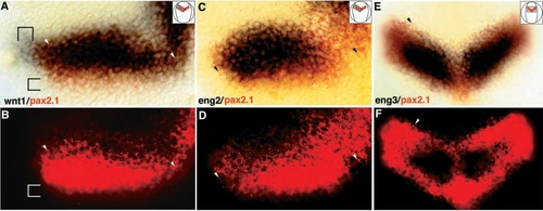
Localization of eng3 and wnt1 (blue) relative to pax2.1 (red/fluorescent) in double in situ hybridizations. All embryos are oriented anterior to the top; insets give the area shown in detail, and arrowheads point to identical landmark cells in corresponding panels. (A) 90% epiboly stage embryo stained for wnt1/pax2.1. The two brackets mark the area where only pax2.1 or wnt1 are expressed. The wnt1 domain extends further laterally than pax2.1, whereas pax2.1 extends further posteriorly; at this stage the anterior boundaries of wnt1 and pax2.1 coincide. (B) Fluorescence image of A. FastRed fluorescence is quenched in the overlapping part by the wnt1 signal, but not posteriorly. (C) 90% epiboly stage embryo stained for eng2/pax2.1. The eng2 domain lies within the pax2.1 domain. (D) The fluorescent image of C, clearly showing that the eng2 cells lie within the pax2.1 domain. (E,F) 1-somite embryo stained for eng3/pax2.1; the initial eng3 expression domain lies within the pax2.1 domain; shortly after, the two domains become coincident (not shown). EXPRESSION / LABELING:
|
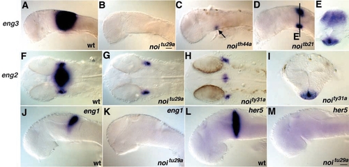
Late marker gene expression during pharyngula stages in noi mutant embryos. Shown are lateral views, anterior to the left (except F-H: dorsal views). (A,B) no eng3 is seen in null mutants at 22 hours, whereas in the strong allele noith44a (C) a ventral eng3-positive spot can still be detected, and both ventral and dorsal expression is visible in weak noitb21 mutants (D,E); E, transverse section. (F-H) 24 hours. eng2 expression in the neural tube is absent in noitu29a; lateral expression in presumptive eye muscles is not affected. In the weak noity31a allele ventral expression is seen, and is located, similar to the eng3 expression, in the ventral neural tube (I; transverse section). (J-M) Eng1 and her5 expression is completely absent at 26 hours. EXPRESSION / LABELING:
|