
- Title
-
Zebrafish hox genes: expression in the hindbrain region of wild-type and mutants of the segmentation gene valentino
- Authors
- Prince, V.E., Moens, C.B., Kimmel, C.B., and Ho, R.K.
- Source
- Full text @ Development

Expression of hox genes in the developing hindbrain; wholemount in situ hybridization with 7 different hox gene riboprobes (purple signal). In most specimens krox-20 was used as a second marker to allow orientation with respect to r3 and r5 (red signal). Embryos were dissected off the yolk and flat-mounted between cover slips for photography. In all panels except E(i) only the hindbrain region is shown, anterior is to the left. Scale bar, 50 μm, where no scale bar shown scale is equivalent to panel above. (A) hoxb1 is expressed in presumptive r4 and posterior from the onset of expression at approximately 90% epiboly, through the 1 somite (1s) stage (i); by the 3s stage (ii) expression in r4 is up-regulated, note r4 lies between the r3 and r5 krox-20 expressing domains (red); hoxb1 expression in r5 and r6 is concomitantly down-regulated. At the 10s stage (iii) the r4 expression domain has sharp borders, abutting the krox-20 expression domains in r3 and r5; more posterior expression is at significantly lower levels in r7 and posterior. At the 20s stage (iv) the same pattern persists, but expression posterior of r4 is no longer apparent in the CNS. (B) hoxa2 is expressed in presumptive r2 and r3 from the onset of expression at the 2s stage (i), the same expression domain persists through the 5s stage (ii). At the 10s stage (iii) high level expression persists in r2 and r3 (note sharp anterior limit of expression at r1/2 boundary); low level expression has spread posteriorly through r4 and r5 (note overlap with krox-20 expression domains). At the 20s stage (iv) neural expression persists in r2 through r5, there are differences in expression levels between the individual rhombomeres such that the highest level is in r2, a slightly reduced level in r3, further reduced in r5 and lowest of all in r4. Neural crest migrating into the 2nd and 3rd branchial arches (arrows) also expresses hoxa2. (C) hoxb2 has an anterior expression limit one rhombomere length more posterior than that of its paralogue, hoxa2, at the r2/3 boundary. The onset of expression is at approximately the 1s stage (i) in presumptive r3 and r5, this was determined in other specimens by colocalization with krox-20; however, to avoid obscuring the weak r5 expression domain the sample shown is a single in situ with only the hoxb2 probe. By the 3-4s stage (ii) the level of r5 expression has increased and there is low level expression in r4 between the two high level domains, in addition there is low level expression posterior to r6. By the 10s stage (iii) expression is confined to rhombomeres 3, 4 and 5, with a step-wise expression gradient from anterior to posterior (highest levels in r3). At the 20s stage (iv) this step-wise gradient persists and expression can also be seen in neural crest cells migrating out into the second branchial arch (arrow). (D) hoxb3 has an onset of expression at the 1-2s stage, within the CNS posterior to r5, by the 3-4s stage (i) expression overlaps the r5 krox-20 domain, the anterior-most expression domain, in presumptive r5 and r6, is at elevated levels compared to more posterior expression. The expression limit and elevated expression levels in r5 and r6 are maintained through the 5s (ii) and 10s (iii) stages. At the 10s stage (iii) neural crest migrating into the 3rd branchial arch from the posterior part of r6, is also expressing hoxb3. By the 30 hour stage (iv) a low level expression domain in r4 has become apparent (arrow). (E) hoxd3 also has an expression onset at the 1-2s stage (i) in the posterior most part of the embryo, by the 3-4s stage (ii) expression has spread rapidly anteriorly to abut the r5 expression domain of krox-20. This r5/6 expression limit is maintained through the 10s stage (iii) and 20s stage (see Fig. 5I). By 30 hours (iv) a more anterior expression domain in a small lateral/ventral group of r5 cells is visible (arrow). (F) hoxx4 has an expression onset at about the 1s stage (i) with an anterior limit lying approximately within r7, expression is at highest levels toward the anterior limit. This expression pattern is maintained through the 5s (ii) and 10s (iii) stages, with the anterior expression limit lying within r7. Even by 30 hours no sharp anterior boundary has been reached (iv). (G) hoxb4 has a similar expression onset to its paralogue, hoxx4, at the 1s stage (i) approximately within r7. This expression domain is maintained at the 5s stage (ii), by the 10s stage (iii) the limit of hoxb4 expression is slightly more anterior than that of hoxx4, reaching the r6/7 boundary by the 15s stage (data not shown). The anterior expression limit is maintained at the r6/7 boundary through 30 hours of development (iv). EXPRESSION / LABELING:
|

Comparison of expression of val (A,B)with hoxb3 (C,D)and hoxb4 (E,F) at the 10s stage (A,C,E) and 20s stage (B,D,F); double in situs with krox-20 (red) to indicate r3 and r5; the precise extent of r5 is indicated with a bracket in A-D. (A) At the 10s stage val is expressed in presumptive r5+r6 (rhombomeres are numbered). (C) There is high level expression of hoxb3 in the corresponding r5+r6 region, note expression in emergent neural crest at the r6 level (arrow). (E) hoxb4 has not reached its anterior expression limit at this stage. (B) At the 20s stage val expression continues to be localized to r5 and r6, there is also expression in the Mauthner neurons in r4 (arrowheads) and low level expression in a small number of emergent neural crest cells (arrow). (D) The high level expression domain of hoxb3 continues to co-localize with the val expression domain in r5 and r6, note expressing neural crest cells (arrow). (F) hoxb4 has now reached its anterior limit at the r6/7 boundary, corresponding to the posterior limit of val expression. EXPRESSION / LABELING:
|
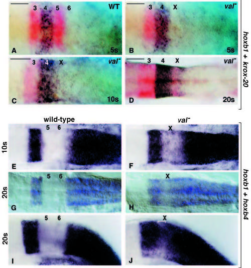
hoxb1 (purple) and krox-20 (red) expression in wild-type (A) and val- (B,C,D) embryos, rhombomeres are numbered. At the 5s stage, there are no obvious differences in the hoxb1 expression pattern between normal (A) and mutant (B) embryos. At the 10s stage the posterior limit of hoxb1 expression in r4 is sharp in wild-type embryos (see Fig. 2Aiii), but not in mutant embryos (C). By the 20s stage there is diffuse hoxb1 staining throughout rX (D). hoxb1 and hoxb4 probes were combined to show expression of both of these genes in wild-type (E,G,I) and val- (F,H,J) embryos. At the 10s stage (E) and the 20s stage (G – dorsal view; I – lateral view) there is a clear zone of nonexpression overlying r5 and r6 in wild-type embryos. In val- embryos at 10s (F) the size of this domain is reduced and some expressing cells are visible within the domain. By the 20s stage (H – dorsal view; J– lateral view) there is expression throughout most of rX, although expression is reduced in dorsal rX in the region where krox-20 expression is expected to localize (indicated by bracket in J). Scale bar, 50 μm. EXPRESSION / LABELING:
|
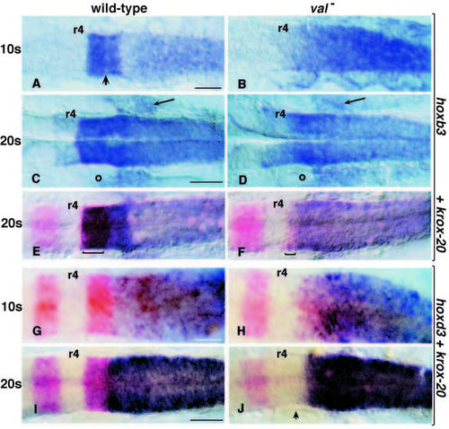
Expression of paralogue group 3 genes in wild-type (A,C,E,G,I) and val- (B,D,F,H,J) embryos, r4 is indicated on each specimen. (A-F) In situ hybridization with hoxb3. At the 10s stage hoxb3 has a high level expression domain in r5+r6 (A, arrow indicates r4/5 boundary), this domain is lacking in the val- embryos (B). By the 20s stage a low level expression domain is apparent in r4 (C,D), high level expression continues in wild-type embryos in r5+r6 (C) next to the otic vesicle (o), the comparable rX expression in val- embryos continues to be at a significantly lower level (D). Neural crest cells migrating posterior to the otic vesicle and expressing hoxb3 are indicated with arrows. E and F show double in situ hybridizations with krox-20 at the 20s stage, to confirm rhombomere locations (the posterior krox-20 expression domains are bracketed in each case). (G-J) In situ hybridization with hoxd3 and krox- 20. In wild-type embryos at 10s (G) and 20s (I) hoxd3 expression reaches the r5/6 boundary to abut the r5 krox- 20 expression domain. In val- embryos at 10s (H) hoxd3 expression is maintained in rX where, similar to hoxb3, it does not overlap with the small rX krox-20 expression domain. At 20s (J) expression is confined to the posterior half of rX, arrowhead indicates the approximate location of the r4/rX interface. Note variable levels of krox20 expression in rX, as discussed in text (compare J with F). Scale bar, 50 μm, where no bar shown, scale is equivalent to adjacent or above panel. EXPRESSION / LABELING:
|

Paralogue group 2 gene expression in wild-type (A,C) and val- (B,D) embryos at the 20s stage reveals a population of hox expressing cranial neural crest migrating posterior to the otic vesicle. In wild-type embryos (A), hoxb2 is expressed in r3, r4 and r5 and in cranial neural crest deriving from r4 and migrating anterior to the otic vesicle (o) into the 2nd branchial arch. In val- embryos (B) the otic vesicle (o) is reduced in size; in addition to the normal population of cranial neural crest migrating anterior of the vesicle, there is a subpopulation possibly deriving from rX, which expresses hoxb2 but migrates posterior to the otic vesicle toward the 3rd arch (arrow). In wild-type embryos (C) hoxa2 is expressed in r2, r3, r4 and r5, and in cranial neural crest migrating into the 2nd and 3rd branchial arches. In val- embryos (D) expression levels are reduced in rX, however there are increased expression levels in neural crest migrating into the 3rd branchial arch (arrow). Scale bar, 50 μm. EXPRESSION / LABELING:
PHENOTYPE:
|