
- Title
-
Pattern formation in janus-mutant zebrafish embryos
- Authors
- Abdelilah, S. and Driever, W.
- Source
- Full text @ Dev. Biol.

Early janus-mutant phenotypes. (A) Wild-type at 16-cell stage. (B) Mild severity of janus-mutant phenotype at 16-cell stage. Separation between the two blastodermal halves is not very pronounced. (C) Strong severity of janus-mutant phenotype at 16-cell stage. (D) Blastoderm disintegration phenotype at the 128-cell stage. Lateral view. Scale bars are 100 μm. |

Gastrulation movements in janus-mutant embryos. (A –D) Wild-type embryo. (E–H) janus-mutant embryo. (A, E) Late blastula-stage embryos prior to gastrulation. (B, F) Dome-stage embryos; blastoderm thinning. (C, D, G, H) Shield-stage embryos; involution/ ingression movements result in the formation of the germ ring within the marginal zone. Convergence movements toward the dorsal side create the shield region (black arrow). (A, B, C, E, F, G) Lateral view with animal pole to the top. (D) Animal view, dorsal toward the right. (H) Lateral–animal view onto one blastoderm, dorsal toward the left. Asterisk indicates the animal pole of the yolk cell, close to the position of the shield. mz, marginal zone; sh, shield. Scale bar is 100 μm. |

ntl expression in shield-stage janus-mutant embryos. (A, D) Wild-type embryo. (B, E) janus-mutant embryo with completely separated blastoderms. (C, F) janus-mutant embryo with partially merged blastoderms lack ntl expression at the animal pole where both marginal zones are fused. (A–C) Animal view. (D–F) Lateral view. mz, marginal zone. Scale bar is 100 μm. |
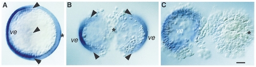
GATA-2 expression at 75% epiboly in janus-mutant embryos. (A) Wild-type embryo. GATA-2 expression marks the nonneural ectoderm in a 180° arc. Boundaries of normal expression are indicated by arrowheads. (B) janus-mutant embryo in which the shield contributes to the two blastoderms. Each half-blastoderm has a ventral GATA-2 expression domain (ve). (C) janus-mutant embryo in which the entire shield is localized to one blastoderm. Only the ventral half-blastoderm has GATA-2 expression. Animal view. (B, C) Separated blastoderms are flattened for documentation. Asterisk indicates position of shield. ve, ventral side. Scale bar is 100 μm. |

Chordamesoderm in janus-mutant embryos at the end of gastrulation. (A,B) Wild-type. The neuroectodermal marker pax[zf-b] (arrowhead) (Krauss et al., 1991) allows to discriminate between ntl and hlx-1 expression domains. (C,D) janus-mutant embryo in which the shield contributes to the two blastoderms. Each shield results in the formation of a complete anterior axis. (E, F) janus-mutant embryo in which the shield is localized to one of the two blastoderms. Only the dorsal blastoderm forms a complete anterior axis. The ventral blastoderm lacks chordamesoderm. (A, C, E) Dorsal view. (B, D, F) Lateral view. Animal pole of the yolk cell is to the top. Hg, hatching gland expression domain of hgg-1; mz, marginal zone; no, notochord expression domain of ntl; pcp, prechordal plate expression domain of hlx-1; tb, tailbud expression domain of ntl. Black dots indicate the marginal zone between the two blastoderms. Scale bar is 100 μm. |

Ventral mesoderm in janus-mutant embryos. (A–C) Diaminofluorene staining of premigratory erythrocytes at the 30-somite stage. (A)Wild-type. (B, C) janus-mutant embryos inwhich the shield was localized to one of the two half-blastoderms have a dorsal half-blastoderm with patches of premigratory erythrozytes. (A, B) Lateral view. (C) Anterior view. bl, erythrozytes; ey, eye. Scale bar is 100 μm. |

Morphogenesis in janus-mutant embryos. The figure comprises the most common phenotypes observed. First three columns: Blastoderms are completely separated at the shield stage. Last two columns: Blastoderms are partially fused at the shield stage. (A –E) Shield is bisected among the two blastoderms. (F–K) Shield is restricted to one of the two blastoderms near the plane of division that bisects among them. (L–P) Shield is restricted to one of the two blastoderms opposite of the division plane that bisects among them. See text for detailed description of morphogenetic events. Lateral views. Asterisks indicate position of shield. ca, complete anterior axis; ia, incomplete anterior axis. Scale bars are 100 μm. |
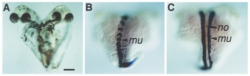
Incomplete axis formations in janus-mutant embryos. (A–C) janus-mutant embryos with complete anterior bifurcation. (A, D) janus-mutant embryos at Day 2 of development. (B) alpha-tropomyosin expression within the incomplete axis addition on the ventral side at the end of somitogenesis. (C) alpha-tropomyosin expression on the dorsal side. (A –C) Lateral view. mu, myotome expression domain of alpha-tropomyosin; no, notochord. Scale bars are 250 μm. |
Reprinted from Developmental Biology, 184(1), Abdelilah, S. and Driever, W., Pattern formation in janus-mutant zebrafish embryos, 70-84, Copyright (1997) with permission from Elsevier. Full text @ Dev. Biol.