- Title
-
Genes establishing dorsoventral pattern formation in the zebrafish embryo: the ventral specifying genes
- Authors
- Mullins, M.C., Hammerschmidt, M., Kane, D.A., Odenthal, J., Brand, M., van Eeden, F.J., Furutani-Seiki, M., Granato, M., Haffter, P., Heisenberg, C.P., Jiang, Y.J., Kelsh, R.N., and Nüsslein-Volhard, C.
- Source
- Full text @ Development
|
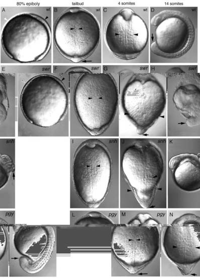
Morphological defects visible in live mutant gastrulae of swr, snh and pgy. Dorsal is to the right in lateral views and animal pole is up in dorsal views. (A-D) Wild type, (E-H) swr (class 5 phenotype), (I-K) snh (class 4 phenotype), (L-N) pg (class 3 phenotype). A lateral view of an 80% epiboly stage wild-type (A) and swr mutant (E) embryo showing the thickened ventral side (white arrowhead) and thinner dorsal axis (black arrowhead) in swr. A dorsal view at bud stage of wild-type (B), swr (F), snh (I), and pgy (L) embryos (the notochord is delineated by arrowheads). Dorsal views of 4- somite stage embryos: wild-type (C), swr (G), snh (J), and pgy (M). The most lateral extent of the somites is marked by arrowheads; an arrow indicates the position of the tailbud, which is not visible in the wild type (C) because it has extended around the yolk. (D,H,K,N) Lateral views of 14-somite stage embryos: wild-type (D), swr (H), snh (K), and pgy (N) (the tailbud is indicated with an arrow). PHENOTYPE:
|

ZFIN is incorporating published figure images and captions as part of an ongoing project. Figures from some publications have not yet been curated, or are not available for display because of copyright restrictions. PHENOTYPE:
|
|
Ntl and myoD expression in swr, snh, pgy and laf mutant embryos. Animal pole or anterior is left or up, except where noted. A dorsal view of whole-mount anti- Ntl antibody stainings at the bud stage of wild type (A); swr (B) where the Ntl notochord domain is about twice as broad as in wild type; snh (C) where the Ntl domain is about 50% broader than in wild type; and pgy (D) where the Ntl domain is about 25% broader than in wild type. In EH, S,T the yolk was removed and then the axis of the embryo flattened onto a slide in order to visualize the entire axis of the embryo (a ‘spread’). Spreads of myoD in situ hybridizations of bud stage embryos in wild type (E,E′), swr (F,F′), snh (G,G′), and pgy (H,H′) mutant embryos. Higher magnifications of the posterior expression (E′-H′) show the mediolateral expansion of myoD expression. Lateral view of a whole-mount myoD staining at the 18- somite stage in wild-type (I) and laf mutant (J) embryos. An arrowhead indicates the enlarged somitic expression of myoD in laf. Dorsal (K,M,Q) and lateral (L,N,R) views of whole-mount myoD stainings of wild-type, swr and snh embryos at the 6-7 somite stage. From a dorsal view of snh (Q) 7 somites are visible. In a lateral view of the same snh embryo (R), it is apparent that only the 5 most posterior somites extend around the circumference of the embryo. Spreads of myoD staining at the 5-6 somite stage in wild type (S) and pgy (T). The wild-type embryo (K) is shown from the vegetal pole (O). In P a vegetal pole view of the swr embryo in M shows the first two somites extending around the circumference of the embryo. The more posterior somites also extend to the ventral side and fuse, but are not in the plane of focus. EXPRESSION / LABELING:
PHENOTYPE:
|
|
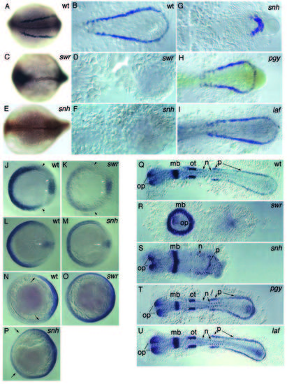
Altered expression of gata1, eve1, fkd3 and pax2 in whole-mount in situ hybridizations of the dorsalized mutants. Expression of gata1 in 8-somite stage wild-type embryos (A,B), swr (C,D), snh (E,F,G), pgy (H), and laf (I) mutant embryos. Double stainings with gata1 and anti-Ntl antibody are shown as whole mounts in A,C,E. Spreads are shown in (B,D,F-I). eve1, gsc double staining at shield stage in wild-type (J) and swr mutant (K) embryos (white arrowheads delineate gsc expression). eve1, gsc double stainings at 70% epiboly in wild-type (L), and snh mutant (M) embryos (white arrow marks gsc expression). fkd3 expression at 70% epiboly in wild-type (N), swr mutant (O), and snh mutant (P) embryos (black arrows indicate the lateral extent of staining). Spreads of pax2-stained 10-somite stage wild type (Q), swr (the most anterior position of the embryo lies in the middle of the ring; R), snh (S), pgy (T), and laf (U) mutant embryos. op, optic vesicle and stalk; mb, midbrain; ot, otic vesicle; n, neuronal and p, pronephric precursor expression domains. EXPRESSION / LABELING:
PHENOTYPE:
|

Unillustrated author statements PHENOTYPE:
|


