- Title
-
Jaw and branchial arch mutants in zebrafish. II. Anterior arches and cartilage differentiation
- Authors
- Piotrowski, T., Schilling, T.F., Brand, M., Jiang, Y.J., Heisenberg, C.P., Beuchle, D., Grandel, H., van Eeden, F.J., Furutani-Seiki, M., Granato, M., Haffter, P., Hammerschmidt, M., Kane, D.A., Kelsh, R.N., Mullins, M.C., Odenthal, J., Warga, R.M., and Nüsslein-Volhard, C.
- Source
- Full text @ Development
|
Photomicrographs and schematic drawings of live (A,B and D) and Alcian blue-stained (C,E,G) wild-type larvae. Color-coded digrams (F,H) of the stained larvae shown in E and G depict distinct cartilaginous elements of the head and pectoral girdle. In H the neurocranium is omitted. C and D present dorsal views, whereas B and G show ventral views of live and stained larvae, respectively. A,E and F show lateral views of these larvae. Abbreviations used throughout all figures: bb, basibranchial; bh, basihyal; ch, ceratohyal; co, coracoid of pectoral girdle; cb, ceratobranchial; c, cleithrum; e, ethmoid plate; ff, facial foramen; hb, hypobranchial; hs, hyosymplectic; m, Meckel’s cartilage; oa, occipital arch; op, opercle; pc, parachordal; pp, pterygoid process of the palatoquadrate; pq, palatoquadrate; t, trabeculae; te, teeth. Color code used in all diagrams: green, Meckel’s; blue, platoquadrate; red, hyosymplectic; yellow, ceratohyal; light blue, interhyal; light green, arches 3-5; grey, neurocranium or ectopic cartilages; white, coracoid of pectoral girdle. Scale bars: A,B,D, 200 μm; C,E-H: 100 μm. |
|
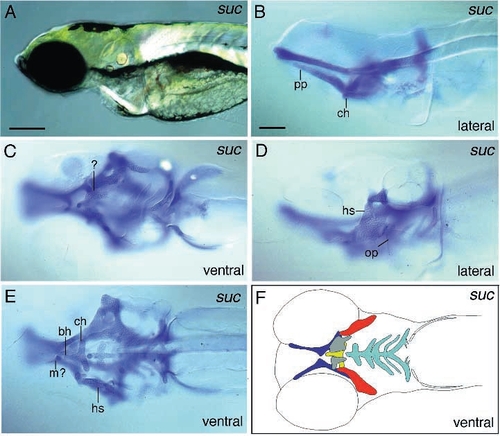
Photomicrographs of lateral views (A,B,D) and ventral views (C,E,F) of sucker mutant embryos reveal that Meckel’s cartilage (lower jaw) is strongly reduced or absent. The diagram (F) represents a composite of elements photographed in C and E. The neurocranium is omitted. The ceratohyal is reduced and fused to the basihyal (E). Although the posterior arches are present, they are reduced. Ventral to the ceratohyal additional unidentified plate-like cartilaginous elements can be detected (C,F). Scale bars: 200 μm (A); 100 μm (B-F). PHENOTYPE:
|
|
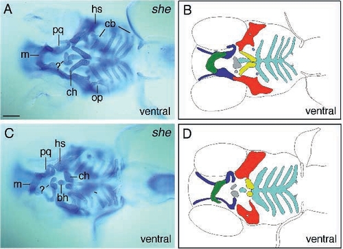
Photomicrographs (A,C) and diagrams (B,D) of ventral views of schmerle (th210) larvae, showing reductions of elements of the first two arches. B and D depict the elements of larvae shown in A and C. Larvae of one egg lay show variable reductions of elements (A,C). In the less-affected larva (A,B) the ceratohyal articulates with the hyosymplectic further to the anterior, but points posteriorly, whereas in the more severely affected specimens (C,D) these elements are reduced to two roundish structures. In both embryos additional pieces of cartilage are located in the vicinity of the basihyal indicated by (?) (C,A). Scale bar, 100 μm. PHENOTYPE:
|
|
Photomicrographs of sturgeon (td204e) (A,B) and hoover (C,D) mutant larvae in which Meckel’s (first arch) is fused with the palatoquadrate. Lateral (A,C) and ventral (B,D) views of stained sturgeon and hoover larvae reveal that the ceratohyal is fused to the hyosymplectic as well. Additional unidentified pieces of cartilage occur in the vicinity of the basihyal and caudal to Meckel’s (A,B,D, arrows). In hoover mutant larvae the ceratohyal cartilage is sometimes divided into two pieces (D). Scale bar, 100 μm. PHENOTYPE:
|
|
Photomicrographs of van gogh (tm208) (A,B), dolphin (C-F) and hanging out (G,H) mutant larvae. In van gogh mutant embryos all elements are severely malformed. A ventral view (A) reveals that Meckel’s is inverted and the ceratohyals are not clearly identifiable any more. The posterior arches are reduced or absent. The ear capsules are misshapen, as can be seen from a dorsal view (A). Live dolphin mutant larvae are characterized by a lack of tissue dorsal to the ethmoid plate (C). The ethmoid plate is arrow-like in shape and consists of fewer chondrocytes anteriorly (E). Lateral (D) and ventral (F) views of stained larvae show that the the ceratohyals fuse dorsally with each other and the overlying basihyal. In larvae homozygous for hanging out, the mouth always stays open (G) and the pharyngeal skeleton points ventrally (H). Scale bars: 200 μm (C and G) and 100 μm (A,B,D,E,F and H). |
|
Photomicrographs of live (A) and Alcian blue-stained (B-F) larvae belonging to the ‘hammerhead-like’ group. The live larvae are characterized by a lack of tissue anterior to the eyes, as can be seen from a dorsal view (A). Hammerhead mutant larvae (A-D) are characterized by a shortened head and kinked elements. All elements are present but consist of smaller and less organized chondrocytes (B-D). The pharyngeal skeleton of pipe tail (ti265) mutant embryos (E,F) is reduced in length but appears thicker, especially the ceratohyal (E). Meckel’s cartilage is located further to the posterior, at about the level of the center of the eyes. The neurocranium is also reduced in length and the ethmoid plate is less differentiated (F). Scale bars: 200 μm (A) and 100 μm (B-F). PHENOTYPE:
|
|
Photomicrographs of several members of the hammerhead-like group stained with Alcian blue. Mutant jellyfish embryos (A,B) can be identified by a severe lack of tissue anterior to the eyes (A, ventral view) and strongly reduced or loss of cartilage. Two elements are always present at the level of the ceratohyals (B, ventral view). In geist (ti240) mutant larvae (C,D) the elements hardly stain with Alcian blue and consist of less organized chondrocytes. Tumor-like outgrowths of chondrocytes are characteristic for knorrig mutant larvae (E,F). From a lateral view (E), additional chondrocytes can be seen, particularly ventral to the ceratohyal (arrow). A ventral view (F) reveals that none of the elements possesses well defined edges. Scale bars: 200 μm (A) and 100 μm (B-F). PHENOTYPE:
|
|
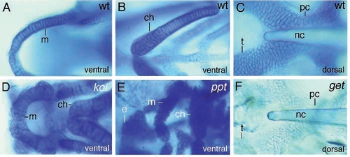
Photomicrographs of cartilaginous elements of wild-type (A-C), knorrig (D), pipe tail (E) and geist (F) mutant larvae. (A,D,E) show ventral close-up views of Meckel’s and the ceratohyal. In knorrig mutant larvae (D), Meckel’s is smaller but thicker and chondrocytes grow along the edges. In pipe tail mutant embryos, Meckel’s lies further to the posterior and all elements consist of smaller but more numerous cells. The ceratohyal is relatively bigger than the other elements. Dorsal views of the neurocranium (C,F) indicate that chondrocytes of geist mutant larvae hardly stain with Alcian blue. |

Unillustrated author statements PHENOTYPE:
|


