
- Title
-
Hematopoietic mutations in the zebrafish
- Authors
- Weinstein, B.M., Schier, A.F., Abdelilah, S., Malicki, J., Solnica-Krezel, L., Stemple, D.L., Stainier, D.Y., Zwartkruis, F., Driever, W., and Fishman, M.C.
- Source
- Full text @ Development
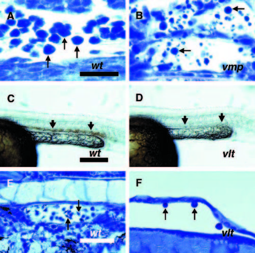
Blood cells are not properly generated during early stages of hematopoiesis in vmp and vlt mutants. Embryos were obtained from crosses between two vmpm62 heterozygotes (A,B) or two vltm651 heterozygotes (C-F). (A,C,E) Phenotypically wild-type embryos. (B,D,F) Mutant siblings. (A,B) Wright-Giemsa stained longitudinal sections from the tails of 27 somite stage (approximately 22.5 hpf) mutant (B) and unaffected sibling (A) embryos. Erythroblasts are seen in both embryos (arrows), but there are many fewer are present in mutants. (C,D) Whole DAF stained 28 somite stage (23 hpf) phenotypically wild-type (C) and mutant (D) embryos. DAF staining is seen in the IMC of wild-type but not mutant embryos (arrows). (E,F) Wright-Giemsa stained longitudinal sections of 2.25 dpf phenotypically wild-type (E) and mutant (F) embryos. Normal blood cells are seen in wild-type embryos, but only abnormal, large cells are seen rarely in mutants (arrows). Scale bars 25 µm (A,B), 200 µm (C,D) and 50 µm (E,F). PHENOTYPE:
|
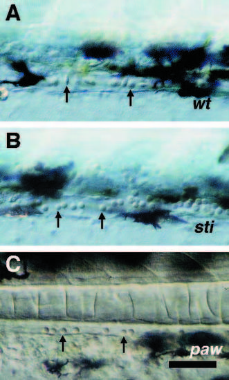
Defects in erythrocyte morphogenesis in sti and paw mutants. DIC photomicroscopy of blood cells (arrows) in tail vessels of zebrafish embryos. (A) Phenotypically wild-type 2.25 dpf embryo from a cross between two stim232 heterozygotes; (B) mutant 2.25 dpf embryo from a cross between two stim232 heterozygotes; (C) mutant 2.25 dpf embryo from a cross between two pawm416 heterozygotes. Scale bar, 50 µm. PHENOTYPE:
|

Defects in erythrocyte morphology and hemoglobin accumulation in clb mutants. Embryos were obtained from a cross between two clbm525 heterozygotes. (A,B) DIC photomicroscopy of blood cells in the caudal arteries of 5 dpf phenotypically wild-type (A) and mutant (B) embryos. (C) Photomicrograph of anterior portions of live 3-day old phenotypically wild-type (top) and mutant (bottom) embryos. The heart appears visibly red (arrow) in the wildtype but not the mutant embryo (arrows). Scale bars, 10 µm (A,B), 400 µm (C). PHENOTYPE:
|

Blood cells are photosensitive in drc mutants. (A-D) DIC videomicroscopy of 2.5 dpf embryos using low-light level illumination and a SIT camera with frame-averaging. All embryos were obtained from a single cross between two drcm87 heterozygotes. Circulation was stopped in the embryos by tricaine overdose. (A) The tail of a phenotypically wild-type embryo raised continuously in room light. Blood cells are seen in the caudal artery (large arrow) and caudal vein (small arrow). (B) The tail of a mutant sibling raised continuously in room light The artery and vein are both present (arrows), but contain no blood cells. (C) Blood cells in the heart of a phenotypically wild-type embryo raised in darkness from blastula stages on. (D) Blood cells in the heart of a mutant sibling embryo raised in darkness from blastula stages on. Abundant blood cells are seen in both the phenotypically wild-type and mutant embryos (arrows). (E) Higher magnification view of blood cells in the caudal artery of a mutant embryo. Blood cells with the typical lentiform morphology for this age are seen (arrows). Scale bar, 100 µm (A-D), 29 µm (E). PHENOTYPE:
|

ZFIN is incorporating published figure images and captions as part of an ongoing project. Figures from some publications have not yet been curated, or are not available for display because of copyright restrictions. |