
- Title
-
The chinless mutation and neural crest cell interactions in zebrafish jaw development
- Authors
- Schilling, T.F., Walker, C., and Kimmel, C.B.
- Source
- Full text @ Development

Embryos homozygous for the chn mutation lack much of the ventral head skeleton and striated musculature. (A,C,E) Wild type; (B,D,F) mutants. (A,B) 30 hour, lateral view. Living wild-type and chnb146 embryos at the prim-15 stage. chn mutants have smaller eyes, a pointed nose and slightly irregular yolks. (C,D) 72 hour, ventral view. By late hatching, much of head skeletal development is blocked. Whole-mounted embryos labelled with Alcian blue. The basilar plate (bp) flanking the anterior notochord is often shorter and thicker than in the wild type. Otoliths within the otic capsule also stain (asterisk). (E,F) 96 hour. Horizontal sections (7 mm) through the pharyngeal region, stained with Mallory’s triple stain for cartilage and muscle (Pantin, 1960). In wild-type embryos, jaw extension moves the mouth opening anterior to the eyes and seven pharyngeal segments differentiate. A mouth opening forms but remains in its original posterior location in chn mutants, surrounded by undifferentiated mesenchyme. All pharyngeal cartilages (blue) as well as striated muscles (red) are absent in chn. Abbreviations: bh, basihyal; cb, ceratobranchials; e, eyes; ep, ethmoid plate; hs, hyosymplectic; mc, Meckel’s cartilage; oa, occipital arch; op, operculum; pbc, posterior basicapsular commissure; p3-6, pharyngeal arches 3-6; pq, palatoquadrate; t, trabeculae cranii. Scale bars, 200 μm. PHENOTYPE:
|
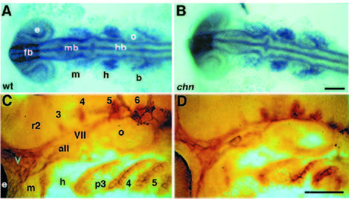
Early neural crest development and the formation of sensory neurons appear unaffected in chn. (A,B) 15 hour, dorsal view. Wholemounted embryos showing dlx2 expression in three major streams of migrating neural crest cells that form the arches. All embryos from a cross between two identified heterozygous chn/+ parents show identical patterns of dlx2 expression. (C,D) 30 hour, lateral view. Pharyngeal arches at prim-15 stage labeled in situ with the monoclonal antibody, zn-5 (Trevarrow et al., 1990). The antibody labels the cell surfaces of segmental clusters of neurons in hindbrain rhombomeres, sensory neurons of the cranial ganglia and endodermal pouches that form between pharyngeal arch primordia. Abbreviations: all, anterior lateral line; b, branchial arches; e, eye; fb, forebrain; h, hyoid arch; hb, hindbrain; m, mandibular arch; mb, midbrain; o, otic vesicle; p3-6, pharyngeal arches 3-6; r2-6, rhombomeres 2-6; V, trigeminal ganglion; VII, facial ganglion. Scale bars, 200 μm. EXPRESSION / LABELING:
|
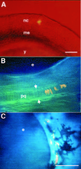
chn neural crest cells do not differentiate as cartilage in the arches. (A) 12 hour, lateral view. A single neural crest cell, labeled with tetramethylrhodamine-dextran is shown after injection. (B,C) 72 hour, lateral view. Computer-combined bright field and fluorescence images. Clones in the mandibular arch in living wild-type (B) and chn mutant (C) embryos, derived from cells located in the largely chondrogenic region of the fate map at early segmentation stages (12-13 hours; Table 1). (B) In the wild type, the clone has contributed several cartilage cells to the palatoquadrate (between arrows). (C) Neural crest cells that migrate into the mandibular arch in chn, remain mesenchymal and never form cartilage although surrounding melanocytes, derived from separate unlabeled neural crest lineages develop normally (Schilling and Kimmel, 1994). Abbreviations: e, eye; me, paraxial mesoderm; nc, neural crest; pq, palatoquadrate; y, yolk. Scale bars, 100 μm. PHENOTYPE:
|

Jaw muscle precursors are specified but do not differentiate in chn. (A,C) Wild-type embryos, (B,D) chn mutant embryos. Whole mounted embryos labelled with anti-Eng antibody were photographed with Nomarski optics. (A) 36 hour, lateral view. In wild type, precursors of two mandibular muscles, the levator arcus palatini and dilator operculi, are present in the mandibular arch and express Eng proteins in their nuclei (Hatta et al., 1990). (B) Cells labelled in a similar location are present in chn mutants (asterisk). (C) 72 hour. At late hatching the Eng-expressing cells have elongated and striated in the wild type. (D) In chn the cells neither elongate nor striate to form contractile muscle fibers. Eng is also expressed in the nervous system at the junction between midbrain and hindbrain in chn mutants as well as wild type. Abbreviations: do, dilator operculi; e, eye; h, hyoid arch; hb, hindbrain; lap, levator arcus palatini; m, mandibular arch; mb, midbrain; o, otic vesicle. Scale bar, 200 μm. EXPRESSION / LABELING:
PHENOTYPE:
|

Mutant neural crest cells form cartilage in the presence of wild-type neighbors. (A) 12 hour, lateral view. Neural crest cells from a donor embryo previously marked with lineage tracer dye are shown after transplantation into the premigratory neural crest of an unlabeled host. A number of labelled cells remain in the suction micropipette. (B) 72 hour, ventral view. In a control, wild-type neural-crest-derived cartilage is identifiable by its morphology in Meckel’s cartilage and the palatoquadrate in a wild-type host. (C) 72 hour, lateral view. A mosaic embryo made by transplanting wild-type neural crest into a chn host. Labelled cartilage cells from the wild-type donor are visible as well as unlabelled cartilage derived from the mutant host (between arrows). (D) 72 hour, ventral view. A mosaic consisting of mutant neural crest transplanted into a wild-type host also shows labelled and unlabelled cartilage. Abbreviations: aa, aortic arch; e, eye; mc, Meckel’s cartilage. Scale bars, 100 μm. |

Rescue of cartilage patterning in chn by transplanted wildtype neural crest cells. Nomarski images of cartilage. 72 hour, ventral view, anterior to the top. (A) Alcian-blue-stained cartilage in the bilateral ceratohyals of a wild-type embryo. (B) A mosaic embryo showing a rod of cells with the stacked appearance of chondrocytes (between arrowheads), extends to the ventral midline in the position of the ceratohyal (asterisk). Abbreviations: ch, ceratohyal; e, eye. Scale bars, 100 μm. |
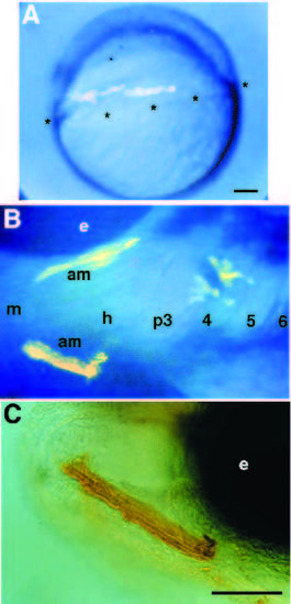
Mutant mesodermal cells form muscles in a wild-type host. All three panels show stages in the development of a single mosaic animal made by transplanting premesodermal cells from a chn donor into a wild-type host at early gastrula stage. (A) 6 hour, lateral view. In the gastrula, rhodamine fluorescence is visible in cells near the equator, at the margin of the blastoderm (asterisks), where they will involute to form mesendoderm. (B) 72 hour, lateral view. Labelled mutant muscle cells are shown in both of the adductor mandibulae muscles in the wild-type mandibular arch of the host. (C) Biotinlabeled, mutant muscle cells in the adductor mandibula. Abbreviations: am, adductor mandibula; e, eye; h, hyoid arch; m, mandibular arch; p3-6, pharyngeal arches 3-6. Scale bars, 100 μm. |