Localization of shh Transcripts in Wild-Type Zebrafish Embryos
Whole embryos were hybridized with digoxygenin-labeled shh riboprobe.
(A and B) Dorsal (A) and lateral (8) views of shh expression at 70% epiboly (the extent of spreading of the blastoderm over the yolk toward the vegetal pole is used as a staging index during epiboly) (Westerfield, 1989). Note the restriction of transcript to the inner cell layer (hypoblast).
(C) Lateral view of an embryo at the 100% epiboly bud stage; by this stage, shh expression extends to the animal pole.
(D) Embryo at 11.5 hr of development. The arrowhead denotes the row of floor plate cells in the neural keel and the underlying notochord cells that express shh.
(E) Embryo at 20 hr of development (22 somites). The arrowhead indicates the notochord that has now switched off shh expression; expression persists at high levels in the floor plate cells (shown in detail in [G]).
(F) Embryo at 26 hr of development (end of somitogenesis). The arrowhead marks shh expression in the telencephalon.
(G) Detail of the trunk region of a 20 hr embryo, similar to that shown in (E), showing shh expression exclusively in the floor plate.
(H and I) Lateral view (H) and top view (I) of an embryo at 33 hr of development, showing shh expression at the posterior end of the pectoral fin bud (arrowhead) and in the first gill (arrow).
(J) Pectoral fin bud of a 33 hr embryo; note that shh expression is confined to the mesoderm and that the superficial epithelial cell layer is devoid of shh expression.
Abbreviations: d, dorsal; eb, epiblast; fp, floor plate; hb, hypoblast; hp, head process; nc, notochord; nk, neural keel; and tb, tail bud.

Comparison of shh Expression with Expression of axial, gsc, and pax-2 in the Head Process of Wild-Type Embryos during Early Embryogenesis
(A, C, and E) At 10 hr of development (end of gastrulation). (B, D, and F) At the five somite stage. (A, C, E, and F) Viewed from the animal pole. (B and D) Lateral views.
(A)shh (horizontal line, marked by arrowheads) and pax-2 (lateral stripes, marked by an asterisk) expression at the end of gastrulation.
(B) shh and pax-2 at the 5 somite stage; pax-2 expression in the posterior midbrain is marked by an asterisk; and the optic stalk is marked by a small arrowhead. shh expression in the tip of the neural keel is shown by an arrow.
(C) axial (arrowheads) and pax-2 (asterisk) expression at the end of gastrulation. Note that the anterior axial expression at this developmental stage compares to the anterior boundary of the shh expression (see [A]).
(D) axial and pax-2 (asterisk) expression at the 5 somite stage. Note that, in contrast with shh, axial expression in the forebrain has now been turned off and extends only to the ventral midbrain/ hypothalamus boundary.
(E) gsc (arrowheads) and pax-2 (asterisk) expression at the end of gastrulation. Note that gsc expression extends further rostral than shh (large arrowhead).
(F) gsc (arrowheads) and pax-2 (asterisk) at the tail bud stage.

EXPRESSION / LABELING:
Gene:
Fish:
Anatomical Terms:
Stage Range: Bud to 5-9 somites

Tissue Sections of Hybridized Embryos Showing shh Expression at Different Developmental Stages
(A-D) Successive transverse sections through the hindbrain of an embryo at the 5 somite stage showing shh expression. (A) Anterior hindbrain.
(B) Hindbrain at the anterior tip of the notochord. (C) Hindbrain at the position of the otic placode. (D) Posterior hindbrain.
(E) Transverse section through the tail of an embryo at the 5 somite stage.
(F) Horizontal section through the spinal cord of an embryo at 26 hr of development. Note the single row of floor plate cells that express shh.
(G and H) Transverse sections through a 48 hr zebrafish embryo. (G) Section through the diencephalon. shh expression is seen in the luminal cells of the foregut and in periventricular cells in the diencephalon (arrowheads). (H) Transverse section at the level of the pectoral fin buds.
(I-M) Serial transverse sections through the head of a 26 hr embryo.
Unless indicated, dorsal is to the top, Abbreviations: d, diencephalon; e, eye; fp, floor plate; ft, foregut; ht, hypothalamus; m, mesencephalon; nc, notochord; nt, neural tube; op, otoc placode; pf, pectoral fin; s, somite; and t, telencephalon.

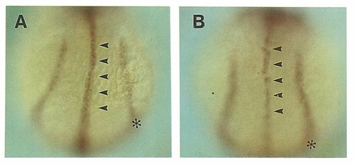
Comparison of shh Expression in ntl Mutant Embryos and Wild-Type Embryos at the Tail Bud Stage
Rostral is to the top. The expression of pax-2 in the Wolffian duct (asterisk) serves as a positional marker. Note the slightly uneven distribution of shh-expressing cells in the notochord precursor cells (arrowheads) of the ntl embryo (see also Halpern et al., 1993) (B) compared with the even rod-like distribution in wild-type embryos (A).

EXPRESSION / LABELING:
Gene:
Fish:
Anatomical Term:
Stage: Bud

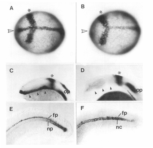
Comparison of shh Expression in cyc Mutant Embryos and Wild-Type Embryos
The right-hand panels show cyc mutant embryos; the left-hand ones show wild-type embryos. Animal pole and lateral views are shown; rostral is to the left. Embryos were simultaneously hybridized with a probe for pax-2 that serves as a positional marker for the posterior midbrain (asterisk).
(A and B) Embryos at the end of gastrulation. Midline shh expression is marked by an arrowhead. Note the absence of the shh transcript in the head process rostral to the pax-2 expression in the mutant embryo (B).
(C and D) Embryos at the 5 somite stage. shh expression in the ventral neural keel is indicated by arrowheads (C); its absence in cyc embryos
(D) is indicated by arrowheads.
(E and F) shh expression in the tail bud at the end of somitogenesis. Note that shh is absent from the ventral CNS of cyc mutant embryos.
Abbreviations: fp, floor plate (in cyc, fp marks the region where the floor plate would normally form); and nc, notochord.

Ectopic Expression of axial after Injection of shh RNA into Fertilized Zebrafish Eggs
(A-C) Whole-mount uninjected and injected embryos hybridized with axial probe after 28 hr of development are shown. (A) axial expression in three wild-type (wt) embryos (left) and three embryos injected (inj) with shh mRNA at the 1- to 4-cell stage (right). Notice the different degree of ectopic axial expression in the head of different embryos, in particular in the ectopic posterior stripe (arrow). (B) axial expression in the head of a control embryo at 28 hr of development. The arrow shows the narrow axial stripe in the diencephalon. The dotted line shows the eye. (C) axial expression in the head of an embryo injected with shh synthetic RNA. Arrowheads mark the broadened axial expression in the diencephalon. The arrow shows the ectopic posterior stripe of axial expression in the midbrain/hindbrain region. The dotted line marks the rudimentary eye.
(D) Horizontal section through the head of a 28 hr wild-type embryo; note the narrow stripe of axial expression through the diencephalon (arrowhead).
(E) Horizontal section through the head of a 28 hr injected embryo; note the broadened stripe of axial expression in the diencephalon (arrowhead) and the ectopic expression in the midbrain (arrow).
(F) Transverse section through the midbrain of a 28 hr injected embryo showing asymmetric distribution of axial-expressing cells.
Abbreviations: d, diencephalon; f, forebrain; h, hindbrain; and m, midbrain.

EXPRESSION / LABELING:
Gene:
Fish:
Anatomical Term:
Stage: Prim-5

Unillustrated author statements

EXPRESSION / LABELING:
Gene:
Fish:
Anatomical Terms:
Stage Range: Bud to Prim-5
Acknowledgments
This image is the copyrighted work of the attributed author or publisher, and ZFIN has permission only to display this image to its users. Additional permissions should be obtained from the applicable author or publisher of the image.

Reprinted from Cell, 75(7), Krauss, S., Concordet, J.P., and Ingham, P.W., A functionally conserved homolog of the Drosophila segment polarity gene hh is expressed in tissues with polarizing activity in zebrafish embryos, 1431-1444, Copyright (1993) with permission from Elsevier. Full text @ Cell