
- Title
-
Expression of truncated Sek-1 receptor tyrosine kinase disrupts the segmental restriction of gene expression in the Xenopus and zebrafish hindbrain
- Authors
- Xu, Q.L., Alldus, G., Holder, N., and Wilkinson, D.G.
- Source
- Full text @ Development
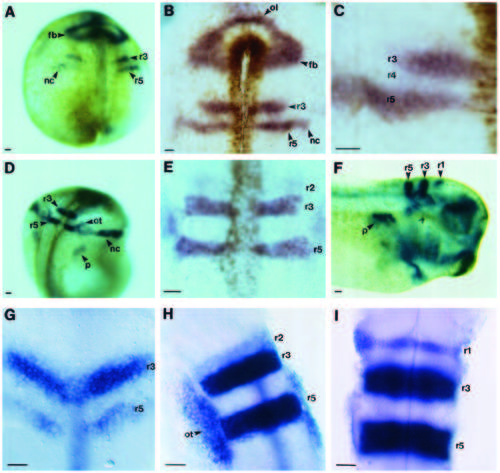
Expression patterns of XSek-1 and rtk1 in the developing hindbrain. (A-F) The expression pattern of XSek-1 during Xenopus development was analysed by whole mount in situ hybridisation. Photographs were taken of either cleared whole embryos (A,D,F), or of the neural epithelium after mounting under a coverslip (B,C,E). (A) Stage 14.5. (B) Rostral neural epithelium at stage 15. (C) Higher magnification view of hindbrain at stage 15. (D) Stage 20. (E) Hindbrain at stage 20. (F) Stage 33. (G-I) The expression pattern of rtk1 during zebrafish development was analysed by whole mount in situ hybridisation. Photographs from a dorsal view were taken after mounting of the hindbrain under a coverslip. (G) 11.5 h. (H) 17 h. (I) 24 h. r, rhombomere; fb, forebrain; nc, neural crest; ol, olfactory placode; ot, otic placode; p, pronephros. Scale bars, 50 µm. EXPRESSION / LABELING:
|
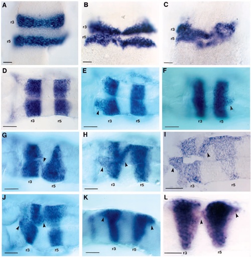
Effect of truncated Sek-1 on krx20 and rtk1 expression in the zebrafish hindbrain. RNA encoding truncated Sek-1 was microinjected into 1 cell of zebrafish embryos at the 2 cell stage. Embryos were allowed to develop to neurula stages, fixed and either krx20 or rtk1 expression analysed by in situ hybridisation. Photographs were taken either of the hindbrain from a dorsal (A-H,J) or lateral view (K,L), or after sectioning in the coronal plane (I). krx20 expression was analysed in (A) uninjected or (B,C) injected embryos fixed at 14 hours of development. rtk1 expression was analysed in uninjected (D) or injected (E-K) embryos analysed at 24 hours of development. (L) krx20 expression in injected 18h embryo. The arrowheads indicate cells expressing krx20 or rtk1 in even-numbered rhombomeres. Scale bars 50 µm. EXPRESSION / LABELING:
|
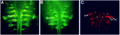
Effect of truncated Sek-1 on reticulospinal neurons in the zebrafish embryo. Reticulospinal neurons were revealed in 3 day zebrafish embryos by retrograde labelling from the spinal cord using LRD (A) Uninjected embryo. (B,C) Embryos injected with RNA encoding truncated Sek-1. The small arrowheads in A and B indicate the locations of corresponding pairs of reticulospinal neurons in r5 and r6. The spacing of these neurons is altered in the injected embryo shown in B. The small arrowheads in C indicate the axons of the duplicated Mauthner neurons in r4. The large arrows labelled ‘m’ indicate the Mauthner neuron. Scale bar, 50 µm. EXPRESSION / LABELING:
|