
- Title
-
Structure of the zebrafish snail1 gene and its expression in wild-type, spadetail and no tail mutant embryos
- Authors
- Thisse, C., Thisse, B., Schilling, T.F., and Postlethwait, J.H.
- Source
- Full text @ Development

Distribution of snail1 RNA during cleavage, blastula and gastrulation. Transcripts from the snail1 gene were revealed by whole-mount in situ hybridization. Lateral views of embryos are oriented with their animal pole up and dorsal side to the right. Animal pole views also have dorsal sides to the right. (A) 2-cell stage, lateral view. Blastomeres are labelled with snail1 antisense probe. ap, animal pole; b, blastomeres; y, yolk cell. (B) Beginning of dome stage, lateral view. The zygotic expression of snail1 has begun on one side of the embryo. Double labelling with the zebrafish goosecoid RNA shows that this is the future dorsal side of the embryo (our unpublished data). (C) Same stage as B, animal pole view. The zygotic RNA occupies an arc on one side of the embryo. The rest of the embryo is labelled more faintly with maternal RNA. (D) 40% epiboly, lateral view. snail1 RNA is localized all around the margin and maternal RNA has mostly disappeared. The two arrows show the position of the margin. (E) 70% epiboly, dorsal view. snail1 transcript disappears from the central part of the embryonic shield (es). (F) 90% epiboly, posterior or vegetal pole view. snail1 RNA is excluded from the axial mesoderm (a) and restricted to the paraxial mesoderm (p) and marginal region. EXPRESSION / LABELING:
|
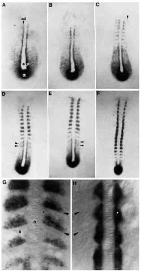
Change in the snail1 expression pattern during somitogenesis. (A) 3-somite stage. snail1 RNA is intensively stained in adaxial cells (ad), a single row of paraxial hypoblast cells (p) adjacent to axial mesoderm (a). Lateral cells are also labelled but in a diffuse manner. The tail bud (tb) is strongly stained. (B) 4-somite stage. Labelling adjacent to the notochord starts to fade posterior to the furrow separating the third and fourth somite. Staining increases just anterior to each newly formed somitic furrow. (C) 7-somite stage. In the segmented mesoderm, signal disappears from adaxial cells. Note the epithelial character of adaxial cells more posteriorly in the unsegmented mesoderm. snail1 RNA is detected anterior to the somitic furrow in a single row of cells. See arrow showing snail1 RNA transiently expressed in the most lateral part of the segmental paraxial hypoblast. (D,E) 10- and 12- somite stages, respectively. In the unsegmented paraxial mesoderm (the segmental plate), there are two stripes of snail1 RNA at segment periodicity posterior to the most recently formed somite (see arrows). As each somite matures, the territory of snail1 expression spreads anteriorly within the somite. (F) 17-somite stage. In the most recently formed somite, snail1 RNA accumulates in a unique line of cells just anterior to the newly formed furrow. (G) Details of snail1 expression pattern in the somite (s) at 17- somite stage, dorsal view. Note the position of the notochord (n) in the middle and the location of the somitic furrow (arrows). snail1 RNA is detected in the posterior compartment of the somite. In the most recently formed somite, a single sheet of cells accumulates snail1 transcript. In older somites, the labelling occupies more of the posterior portion of the somite. The star indicates the position of muscle pioneer precursors which are not labelled with snail1 RNA. (H) 17-somite stage. Dorsal view of an embryo probed with a-tropomyosin. The arrow indicates the position of the somitic furrow. atropomyosin is detected in adaxial cells and in particular, in muscle pioneer precursors (indicated by a star). Note morphogenetic changes in a-tropomyosin-expressing cells as somites mature. EXPRESSION / LABELING:
|
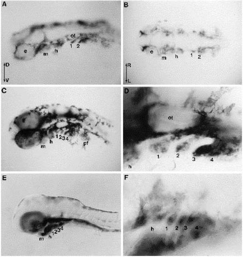
Late expression pattern of snail1 in the head. Embryos were dissected and the yolk was removed. (A) 24 hour embryo, dorsolateral view and (B) 24 hour embryo, dorsal view. snail1 RNA is localized in various cells around the eyes (e) and otic capsule (ot), and in pharyngeal arch primordia (mandibular arch (m), hyoid arch (h), and the first (1) and second (2) gill segments). D, Dorsal; V, ventral; R, right; L, left. (C) 36 hour embryo, lateral view. The extent of labelling has expanded in the branchial arches and is easily distinguishable in the mandibular (m) arch and the hyoid (h) arch, and in the first four (1, 2, 3 and 4) gill arches. Pectoral fin buds (pf) also accumulate snail1 transcript. (D) High magnification of the ear region (ot, otic capsule) and caudal branchial arches focusing on snail1 RNA expression in gill arches. The mesenchyme of each arch is composed of neural crest cells and paraxial mesodermal cells. (E) 60 hour embryo, lateral view. snail1 RNA disappears from the middle of the arches where chondrocytes are beginning to differentiate. (F) Details of the embryo shown in E, focusing on branchial arches. |
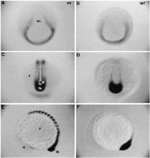
Expression pattern of snail1 in spadetail embryos. (A-D) Posterior views; (E,F) lateral views. (A,B) 80% epiboly; (C,D) 10-somite stage; (E,F) 12-somite stage. (A,C,E) Wild-type embryos; (B,D,F) spadetail embryos. While snail1 RNA lines the lateral border of the embryonic shield in wild-type (A), it is missing in spadetail embryos (B). The axial mesoderm is broader in a spadetail embryo (C) than in a wild-type embryo (D). Wild-type embryos have extensive areas containing snail1 transcripts in the paraxial mesoderm and somites (E), while spadetail embryos have much less snail1 transcript in the paraxial region (F). In the mutant, snail1 transcript is present only in the tail bud and in a few cells scattered along the axial mesoderm. EXPRESSION / LABELING:
|

Expression pattern of snail1 in no tail embryos. (A,B) Posterior views of 5-somite stage; (C,D) lateral views of 12-somite stage; (A,C) wild-type embryos; (B,D) no tail embryos. Compared to a wild-type embryo (A), note the reduction of snail1 signal in no tail, especially in the tail bud, even though this no tail embryo was stained for 6 hours to be able to see a clear signal, and the wild-type control was stained for just 2 hours. The axial mesoderm territory is broader in no tail embryos. In contrast to wild type (C), an overstained no tail embryo shows an absence of snail1 staining in the tail. Since the mutant embryo was stained for a long time, background staining started to become visible. EXPRESSION / LABELING:
|