- Title
-
Zebrafish as a Model to Evaluate a CRISPR/Cas9-Based Exon Excision Approach as a Future Treatment Option for EYS-Associated Retinitis Pigmentosa
- Authors
- Schellens, R., de Vrieze, E., Graave, P., Broekman, S., Nagel-Wolfrum, K., Peters, T., Kremer, H., Collin, R.W.J., van Wijk, E.
- Source
- Full text @ Int. J. Mol. Sci.
|
Schematic representation of the domain architecture of human and zebrafish EYS proteins. Both human and zebrafish EYS proteins are comprised of a repetitive protein domain architecture that includes epidermal growth factor (EGF) domains, EGF-like domains and laminin G (LamG) domains. Skipping of eys exon 40-44 results in the exclusion of one LamG and two EGF domains, indicated with a dashed boxed. Numbers indicate amino acids. |
|
Eys localization in adult zebrafish photoreceptor cells. Immunoelectron microscopy images of adult zebrafish retinas show that Eys localizes at (A,B) the connecting cilium, periciliary membrane, cone accessory outer segments, and (C,D) photoreceptor ribbon synapses. CC: connecting cilium, PCM: periciliary membrane, AOS: accessory outer segment, IS: inner segment, OS: outer segment. EXPRESSION / LABELING:
|
|
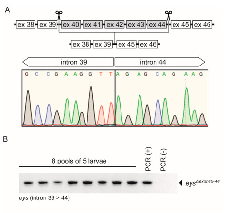
Characterization of the stable eysΔexon40-44 zebrafish line. (A) Schematic representation of exon excision approach (upper panel). Sanger sequencing confirmed the presence of the anticipated exons 40-44 excision in injected embryos (1 day post fertilization (dpf)); lower panel). (B) PCR analysis identified germline transmission in 8 out of 8 pools of 1 dpf embryos obtained by breeding an injected adult with a wild-type animal. PCR (+): positive PCR control, PCR (-): negative PCR control. |
|
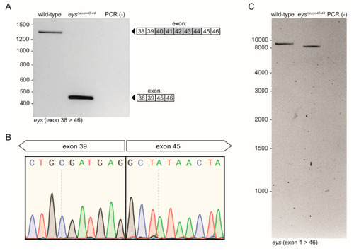
Transcript analysis of EXPRESSION / LABELING:
|
|
Immunohistochemistry on retinal sections of wild-type, |
|
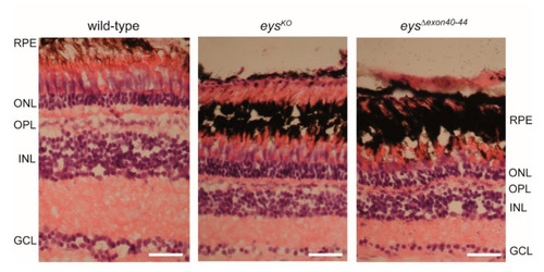
Histological examination of wild-type, eysKO and eysΔexon40-44 zebrafish retinas. Light microscopy of retinal sections from adult zebrafish (15 months post fertilization (mpf)) stained with hematoxylin (purple) and eosin (red). Retinas of the eysΔexon40-44 and eysKO lines are morphologically indistinguishable. In both the eysΔexon40-44 and eysKO retinas, a reduction of the thickness of all retinal layers was observed as compared to wild-type controls. In addition, photoreceptor outer segments seem to be shortened and disorganized in both eysΔexon40-44 and eysKO as compared to wild-type retinas. Scale bar: 20 μm. RPE: retinal pigment epithelium, ONL: outer nuclear layer, OPL: outer plexiform layer, INL: inner nuclear layer, GCL: ganglion cell layer. |
|
Visual Motor Responses in wild-type, eysKO and eysΔexon40-44 zebrafish. (A) A representative eye-specific Light-ON Visual Motor Response (VMR) from a single trial, presented as the distance moved (mm) per second, is shown for the time frame of 15 s prior to and after light alternation. Note that the response to light is less pronounced in the eysKO and eysΔexon40-44 larvae as compared to the strain- and age-matched wild-type larvae (5 dpf; n = 16 per group). (B) The distance moved (mm, mean of 4 Light-ON transitions per larva) during the first second after the Light-ON transition for wild-type, eysKO and eysΔexon40-44 zebrafish. The distance moved was significantly lower in the eysΔexon40-44 larvae (2.04 mm ± 0.66 (mean ± SD, n = 48)) than in wild-type larvae (2.72 mm ± 0.93 (mean ± SD, n = 48)), while no significant difference was observed between the eysKO larvae (1.75 mm ± 0.57 (mean ± SD, n = 47)) and eysΔexon40-44 larvae (5 dpf; n= 47 larvae). *** indicates p < 0.0001. Trials were conducted as 3 biological replicates containing all genotypes in each trial. PHENOTYPE:
|


