FIGURE SUMMARY
Title

Loss of Gap Junction Delta-2 (GJD2) gene orthologs leads to refractive error in zebrafish

Authors
Quint, W.H., Tadema, K.C.D., de Vrieze, E., Lukowicz, R.M., Broekman, S., Winkelman, B.H.J., Hoevenaars, M., de Gruiter, H.M., van Wijk, E., Schaeffel, F., Meester-Smoor, M., Miller, A.C., Willemsen, R., Klaver, C.C.W., Iglesias, A.I.
Source
Full text @ Commun Biol

Loss of Cx35.5 (<italic>gjd2a</italic>) leads to reduced ocular dimensions.

SD-OCT recordings of size-matched juvenile-to-adult zebrafish indicating the temporal dynamics of the eye. All ocular metrics were corrected for the tissue-specific refractive index. a Single B-scan image of a 3 mpf zebrafish eye. The area is defined as the axial length that spans from the apical part of the corneal epithelium to the anterior border of the RPE (green line). The RPE is represented by the hyperreflective melanin-rich band (magenta), of which the anterior part comprises a sharp-cut border, used as a posterior landmark for the axial length. The gradient refractive index of the spherical zebrafish lens was used as a correction factor to acquire this image (see “Methods”), and the brightness was enhanced for better visualization of the transparent lens. Individual compartments: cornea (light blue), lens (red), vitreous chamber (yellow), neural retina (orange). b, c Axial length of gjd2a (Cx35.5) (b) and gjd2b (Cx35.1) (c) mutant eyes of juvenile (1.5–2 mpf) and adult zebrafish (3 mpf). b The gjd2a (Cx35.5) eyes were significant reduced in axial length compared with WT at 1.5 mpf (effect size = −47 µm, p < 0.001), 2 mpf (Effect size = −48 µm, p < 0.001), and 3 mpf (effect size = −43 µm, p < 0.001). c The gjd2b (Cx35.1) mutant eyes showed no significant alteration relative to WT. d, e, f Dimensions of significantly altered ocular compartments in gjd2a (Cx35.5) mutant eyes. See Supplementary Data S1 and S2 for a full statistical report and dimensions of individual compartments. Sample size: n = 40 eyes for each genotype and time point. Error bars: SEM. Significance: ns = not significant, *p < 0.05, **p < 0.01, ***p < 0.001. Scale bar: 100 µm. Mpf months post-fertilization, SD-OCT spectral-domain optical coherence tomography, RPE retina pigmented epithelium.

Opposite refractive error in <italic>gjd2a</italic> (Cx35.5) and <italic>gjd2b</italic> (Cx35.1) mutants.

a Schematic illustration of the eccentric photorefractor setup. b Calibration by −6 diopter (D), −2D, 0D, +4D, and +10D lenses led to a conversion factor of 1.924 (R2 = 0.971). c Typical intensity profile of a hyperopic (asterisk) 3mpf gjd2a (Cx35.5) mutant and myopic (arrowhead) lrp2 mutant. The gjd2b (Cx35.1) mutant shows both myopic (arrowhead) and hyperopic (asterisk) contralateral features. d, e RE in the gjd2a (Cx35.5) (d) and gjd2b (Cx35.1) (e) mutants at 1.5 mpf, 2 mpf, 3 mpf, and 9 mpf. d Loss of Cx35.5 (gjd2a) results in a significant (p < 0.001) and progressive hyperopic shift in refractive status. e Loss of Cx35.1 (gjd2b) is linked to a significant (p < 0.001) and progressive myopic shift. f Mutant refractive status normalized against the baseline refraction of WT controls, indicated by the relative RE. Sample size: n = 20 eyes for each genotype and age. Error bars: SEM. Significance: ns = not significant, *p < 0.05, **p < 0.01, ***p < 0.001. Scale bars: 50 µm (c). RE refractive error.

PHENOTYPE:
Fish:
Observed In:
Stage Range: Days 45-89 to Adult

Nuclear cataract in <italic>gjd2b</italic> (Cx35.1) mutants.

a, b Coronal SD-OCT sections of typical 3 mpf lenses of WT control (a) and gjd2b (Cx35.1) mutant (b) fish. c Proportion of cataractous lenses in 1.5 mpf, 2 mpf, and 3 mpf SD-OCT data (n = 40 eyes) indicating an increasing prevalence in gjd2b (Cx35.1) mutants. dg DIC microscopy of 6 mpf fish lenses allows visualization of opaque nuclear lens fibers in lenses of gjd2b (Cx35.1) mutants (e) and (g) and transparent WT control lenses (d) and (f). d, e DIC 20× magnification (f) and (g) 40× magnification. h Proportion of cataractous lenses by SD-OCT and DIC microscopy at 6 mpf (n = 24 eyes). i Ratio of opaque pixels: total pixels indicating the opacity of 6 mpf coronal SD-OCT sections (n = 24 eyes). Error bars: SEM. Significance: ns = not significant, *p < 0.05, **p < 0.01, ***p < 0.001. DIC microscopy: differential interference contrast microscopy.

PHENOTYPE:
Fish:
Observed In:
Stage Range: Days 45-89 to Adult

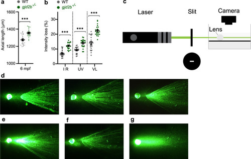
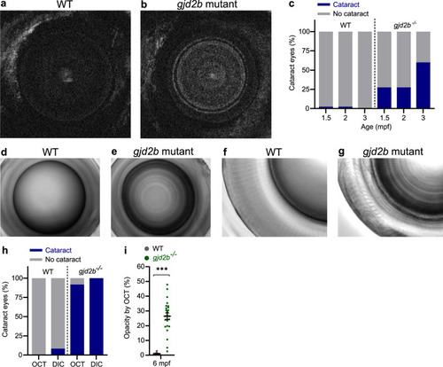
a SD-OCT shows a significant increase in axial length in 6 mpf gjd2b (Cx35.1) mutants (n = 24 eyes). b Loss of lenticular projection intensity of isolated mutant lenses (n = 24 eyes), the following wavelengths were used: IR light (940 nm), UV light (365 nm), and broad-spectrum visible light (380–760 nm). c Schematic illustration of the ex vivo lenticular light propagation setup. d Examples of light propagation in three isolated 6 mpf WT control lenses. e–g Examples of light scattering and multifocality by two moderately cataractous lenses (e) and (f) and light diffusion by a severely cataractous lens (g) of the 6 mpf gjd2b (Cx35.1) mutant. Error bars: SEM. Significance: ns = not significant, *p < 0.05, **p < 0.01, ***p < 0.001. IR infrared, UV ultraviolet.

B-wave potential and spatial acuity in <italic>gjd2a</italic> (Cx35.5) and <italic>gjd2b</italic> (Cx35.1) mutants.

a Electroretinogram showing the average B-wave potential for eyes of gjd2a (Cx35.5) mutants, gjd2b (Cx35.1) mutants, and WT control fish at 2.5 mpf (n = 22 fish). b Maximum B-wave amplitude response. c Spatial visual acuity indicated by the proportion of fish completing a minimum of three subsequent optokinetic responses at spatial frequencies ranging from 0.15 to 0.40 cpd. d Nasally directed ETMs per 10-s interval for spatial frequencies ranging from 0.15 to 0.25 cpd. Error bars: SEM. Significance: ns = not significant, *p < 0.05, **p < 0.01, ***p < 0.001. ETM eye-tracking movements, cpd cycles per degree.

Ocular expression of Cx35.5 (<italic>gjd2a</italic>) and Cx35.1 (<italic>gjd2b</italic>).

Immunostaining showing the topographic distribution of Cx35.5 (gjd2a) and Cx35.1 (gjd2b) throughout the 2.5 mpf zebrafish retina (a)–(t) and Cx35.1 (gjd2b) throughout the 6 mpf zebrafish lens (u). The retinal sections in the left and right column are stained for respectively anti-pan-Cx35 and anti-Cx35.5 (gjd2a) (both in red) and each row indicates the genotype. DAPI (blue) and anti-actin (green) are used for orientation. The staining in the WT retina for anti-pan-Cx35 (a)–(d) and the specific anti-Cx35.5 (e)–(h) reveal generally overlapping patterns of localization in the IPL, OPL, and photoreceptor layer. In the gjd2a (Cx35.5) mutants the anti-pan-Cx35 staining (i)–(l) supports that this antibody recognizes both Cx35.5 (gjd2a) and Cx35.1 (gjd2b) and reveals the localization of Cx35.1 (gjd2b). The absence of staining in the gjd2a (Cx35.5) mutants with the anti-Cx35.5 antibody (m)–(p) support the specificity of the antibody and confirms the Cx35.5 (gjd2a) localization presented in (e)–(h). The presence of staining in the gjd2b (Cx35.1) mutants with the anti-pan-Cx35 antibody (q)–(t) support that this antibody recognizes both Cx35.5 (gjd2a) and Cx35.1 (gjd2b) and supports the localization of Cx35.5 (gjd2a). u Anti-pan-Cx35 immunostaining in isolated 6mpf WT and Cx35.1 (gjd2b) mutant lenses. A modest lenticular appearance at the outer cortical layer can be observed in WT control while absent in gjd2b (Cx35.1) null mutants. The gjd2b (Cx35.1) null mutant lens shows a nuclear ring structure (arrows). Scale bars: 5 µm (a)–(t), 100 µm (u). IPL inner plexiform layer, OPL outer plexiform layer.

Single-cell transcriptome analysis of larval WT zebrafish.

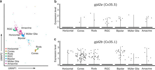
Single-cell transcriptome analysis reveals gjd2a (Cx35.5) and gjd2b (Cx35.1) expression in a wide variety of retinal cell types. a Annotated retinal cell clusters isolated from a single-cell RNA-seq data set of 2–5 dpf larval zebrafish. b, c Expression of gjd2a (Cx35.5) (b) and gjd2b (Cx35.1) (c) in seven identified retinal cell clusters. Each dot represents an individual cell and its expression level of the indicated gene. UMAP uniform manifold approximation and projection, RGC retinal ganglion cell.

Gap junction regulators and the effect on axial growth.

Simplified illustration summarizing the relation between (un)coupling of retinal gap junctions and axial eye growth. We hypothesize, based on reported literature and the outcome of this study (asterisk), that the (un)coupling of retinal gap junctions may play an intermediate role in controlling emmetropization. The exact mechanism of how Cx35/Cx36 uncoupling leads to reduce axial eye growth is unclear (dotted line arrow).

ZFIN is incorporating published figure images and captions as part of an ongoing project. Figures from some publications have not yet been curated, or are not available for display because of copyright restrictions.

Acknowledgments
This image is the copyrighted work of the attributed author or publisher, and ZFIN has permission only to display this image to its users. Additional permissions should be obtained from the applicable author or publisher of the image. Full text @ Commun Biol