
- Title
-
Cells with Many Talents: Lymphatic Endothelial Cells in the Brain Meninges
- Authors
- Suárez, I., Schulte-Merker, S.
- Source
- Full text @ Cells
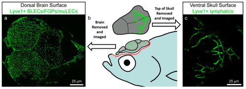
Zebrafish brain lymphatic endothelial cells (BLECs)/Fluorescent Granular Perithelial cells (FGPs)/mural lymphatic endothelial cells (muLECs) and meningeal lymphatic vessels in adult specimens. Image modified from Castranova et al. [49]. (a) Dorsal confocal image showing BLECs/FGPs/muLECs extended over the whole brain surface. (b) Schematic representation of an adult zebrafish depicting the dissection procedure for imaging of BLECs/FGPs/muLECs and intracranial meningeal lymphatic vessels. (c) Confocal image of the inner surface of the skull showing the meningeal lymphatic vessels that stay attached to the skull after dissection. |

Zebrafish BLECs in embryonic and larval specimens. (a,b) Schematic representations of zebrafish showing the same orientation and close-up regions (blue squares) as in c1 (a) and c2-c4 (b). (c) Confocal images from van Lessen et al. [50]. Lympho-venous structures are depicted in green and blood vessels in red. BLECs (arrow heads) start sprouting at 56 hpf from behind the PHS and migrate along the MsV (c1). By 5 dpf they have formed a bilateral loop in the TeO (c2). Cells in the loop are able to take up injected dyes such as pHrodo (red) and IgG-Alexa647 (blue) (c3). BLECs keep spreading above the whole brain area (c4), where they will stay as single cells throughout the lifespan of the animal. Dpf, days post fertilization; hpf, hours post fertilization; MsV, mesencephalic vein; PHS, primary head sinus; TeO, optic tectum. |

Murine leptomeninges contain cells expressing BLEC markers. Image adapted from Shibata et al. [55]. (a) and (c) Coronal sections of a zebrafish and a mouse brain, respectively, equivalent to the imaging areas in (b,d). To facilitate comparison, (b,d) are both displayed with the parenchyma at the bottom and the meninges at the top. (b) Zebrafish BLECs (white arrows) express Vegfr3 (green) and are located adjacent to a meningeal blood vessel (red). The area of the meninges is marked with a white bracket. DAPI (blue) highlights the nuclei. (c) IHC of a 17 week old mouse reveals the presence of VEGFR3 expressing cells (white arrows) adjacent to meningeal blood vessels (magenta). These cells are limited to the region of the meninges (white bracket) and do not penetrate into the brain parenchyma. NG2 (red) labels pericytes and smooth muscle cells, Hoechst (blue) marks nuclei. (e–e’’’) IHC of mouse brain section showing that the cells within the meningeal layers express the BLEC markers MRC1, LYVE1 and VEGFR3. |