FIGURE SUMMARY
Title

The SARS-CoV-2 receptor and other key components of the Renin-Angiotensin-Aldosterone System related to COVID-19 are expressed in enterocytes in larval zebrafish

Authors
Postlethwait, J.H., Massaquoi, M.S., Farnsworth, D.R., Yan, Y.L., Guillemin, K., Miller, A.C.
Source
Full text @ Biol. Open

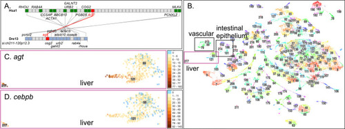
(A) The section of H. sapiens chromosome 1 (Hsa1) that contains AGT is conserved with the segment of D. rerio chromosome 13 (Dre13) that contains agt. (B) The 220 clusters from the zebrafish scRNA-seq Atlas (Farnsworth et al., 2020). Boxes indicate the liver, vascular endothelium, and intestinal epithelium clusters. (C) Cells in hepatocyte clusters c217, c121, and c121 express agt. Each dot represents a cell. Color intensity indicates expression level according to the scale at the right [the number of Unique Molecular Identifiers (UMIs; unique reads) in each individual cell that mapped to the gene of interest]. Blue cells are not expressing. (D) Expression of the Agt-regulator cebpb in larval liver cells.

Evolution of Angiotensin sequences. Arrows indicate human and zebrafish sequences. Species organized according to published phylogenies (Hughes et al., 2018; Upham et al., 2019). Ang I and Ang II sequences indicated at bottom. Table S1 contains Latin names and accession numbers.

Genomics and expression of ace, agtr, and nr3c2. (A) Conserved syntenies verify orthology of zebrafish ace to human ACE. (B) Expression of ace in a specific intestinal epithelial cell type in c152. (C) Clusters c101, c152, c168, and c214 are intestinal epithelial cell types (Farnsworth et al., 2020). (D) The age of each cell, as indicated in the insert (two replicates at each of three developmental stages), showed that ace is expressed in a 5 dpf intestinal cell type c152 that does not exist at 1dpf or 2dpf. (E) Double conserved synteny of zebrafish agtr1a and agtr1b to human AGTR1. (F) agtr1b was expressed only in endothelial cells (c88). Expression of agtr1a was not detected. (G) Conserved synteny of agtr2 and AGTR2 confirms orthology. (H) Expression of agtr2 was detected in c88 and c79 endothelial cells, including some in which agtr1b was detected. (I) Conserved syntenies for the aldosterone receptor gene nr3c2. (J) Expression detected for nr3c2 in intestinal epithelial clusters c168 and c101. (K-L) Expression of nr3c2 in the embryonic periderm (c0, c77) and in larval gills (c5).

Genomics and expression of ace2, enpep, anpep and dpp4. (A) Conserved synteny supports orthology of human ACE2 to zebrafish ace2. Expression of ace (B), ace2 (C), enpep (D), anpepb (ENSDARG00000041083) (E), anpeplb.1 (ENSDARG00000103878) (F), and dpp4 (G). All were expressed mainly in the intestinal epithelial cell type c152.

Conserved syntenies and expression of SLC6A19-related genes. (A) Conserved syntenies show double conserved synteny of a part of Dre16 containing slc6a19b and a portion of Dre19 containing slc6a19a.1, and slc6a19.2 and their relationship to human chromosome Hsa5 around SLC6A19. (B-D) Expression of slc6a19a.1, slc6a19a.2, and slc6a19b, respectively, in the zebrafish Atlas in c152 and c168, which represent the same larval intestinal epithelial cells that expressed ace and ace2.

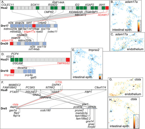
(A) Double conserved synteny between Dre17 and Dre20 confirming co-orthology of adam17a and adam17b to ADAM17 and their origin in the TGD. (B) Expression of adam17a in intestinal epithelium and (C) vascular endothelium. (D) Conserved syntenies verify orthology of tmprss2 to TMPRSS2. (E) Expression of tmprss2 was detected in one cell in intestinal epithelium c168 and in only 12 other cells broadly dispersed in the atlas. (F) Human CTSL shares conserved syntenies with zebrafish ctsla. (G) Expression of ctsla in the endothelium and (H) intestinal epithelium.

Genomics and expression of Apelin and its receptors. (A) Phylogenetic tree (ENSGT01000000214406) with the APLNR/’aplnr2’ (suggested new name aplnr, ENSDARG00000004447) subtree (blue in panel B) expanded. (B) The same tree with the ‘aplnra’/’aplnrb’ (suggested new name aplnrla, ENSDARG00000002172)/aplnrlb (ENSDARG00000036670) subtree (orange in panel A) expanded. (C) A dot plot representing orthologs and paralogs of Hsa11 genes versus zebrafish (Dre) chromosomes plotted directly above the location of each Hsa11 gene showing extensive conservation with Dre7, the site of aplnr, but not Dre8 or Dre10, the locations of aplnrla and aplnrlb, respectively. (D) A dot plot of chicken chromosome Gga15 versus zebrafish chromosomes showing the sites of aplnrla and aplnrlb and their ortholog in chicken (Aplnrl, ENSGALG00000047717) identified in the tree in B. (E-H) Expression of apln, aplnr, aplnrla, and aplnrlb in endothelial cells.

Identifying the ace2-expressing cell type. (A-K) In situ hybridization for ace2 and slc6a19a.1. (A-G) 5 dpf larvae, (A) lateral view and (B) ventral view of ace2 expression. (C) Cross-section of larva stained for ace2 expression at level of the dotted line in B. (D) Close-up of the middle gut cross-section showing intestinal epithelial cells expressing ace2. (E) Lateral view and (F) ventral view of slc6a19a.1 expression. (G) Cross-section at level of the dotted line in F. (H-K) Adults. H, ace2 expression in middle gut, with (J) no expression in kidney. (I,K) slc6a19a.1 expression in middle gut and kidney, respectively. (L-P) Feature plots of intestinal epithelial cells expressing ace2 and marker genes for intestinal stem cells (mki16), enterocytes (villin, vil1), enteroendocrine cells (anxa4), and goblet cells (agr2). (Q) Umap plot for subclustering of cells in original c152, c168, and c214. (R) Violin plot for ace2 in the subclusters. (S-V) Violin plots for genes representing regions along the mouse intestine, including (S) duodenum (ada); (T) jejunum (fabp2); (U) ileum (fabp6); and (V) colon (lamp2). dg, distal gut; e, eye; ib, intestinal bulb; iec, intestinal epithelial cell; k, kidney; kt, kidney tubule; mg, middle gut; n, notochord; nt, neural tube; s, somite. Scale bar in A for A, B, E, F, 100 μm; in C, 50 μm; in D for D and G, 50 μm; in I for H-K, 100 µm.

Acknowledgments
This image is the copyrighted work of the attributed author or publisher, and ZFIN has permission only to display this image to its users. Additional permissions should be obtained from the applicable author or publisher of the image. Full text @ Biol. Open