FIGURE SUMMARY
Title

Bmp8a is an essential positive regulator of antiviral immunity in zebrafish

Authors
Zhong, S., Li, H., Wang, Y.S., Wang, Y., Ji, G., Li, H.Y., Zhang, S., Liu, Z.
Source
Full text @ Commun Biol

Bmp8a inhibits RNA virus replication both in vitro and in vivo.

a, b ZFL cells were transfected with bmp8a (1 μg) or empty vector (1 μg), respectively. The cells were infected with GCRV (5 × 10⁴ TCID50 per ml) at 24 h of post-transfection, and the culture supernatants were collected at 72 h of post-infection. The cell monolayers were fixed with 4% paraformaldehyde for 1 h and stained with 0.5% crystal violet for 2 h (a), and the viral titers of the supernatants were determined by TCID50 assays (b). c, d Similar as (a, b) but in EPC cells. e, f ZFL cells were treated with rBmp8a (final concentrations of 5 μg/ml) or PBS co-incubation with GCRV (5 × 10⁴ TCID50 per ml), and the culture supernatants were collected at 72 h of post-infection. The cell monolayers were fixed with 4% PFA for 1 h and stained with 0.5% crystal violet for 2 h (e), and the viral titers of the supernatants were determined by TCID50 assays (f). g, h Similar as (e, f) but in EPC cells. i The expression of GCRV RNA in the liver, kidney, intestine, and spleen from wild-type (WT) or bmp8a−/− zebrafish injected i.p. with 50 µl of GCRV (108TCID50 per ml) for 72 h. j, k Kaplan–Meier analysis of the overall survival of WT (n = 20) or bmp8a−/− zebrafish (n = 20) which were injected i.p. with 50 µl of GCRV (108TCID50 per ml) or SVCV (108TCID50 per ml) and monitored every 12 h after infection. l Kaplan–Meier analysis of the overall survival of WT (n = 8) or bmp8a−/− zebrafish (n = 8) which were injected i.p. with 50 µl of TSVDV (crude virus extracts filtered by a 0.45 μm microporous membrane) and monitored every 12 h after infection. The expression of zebrafish actb1 was used as an internal control for the qRT-PCR. Data were from three independent experiments (ai) or two independent experiments (jl). Data were analyzed by Student’s t-test (two-tailed) or log-rank (Mantel–Cox) test and were presented as mean ± SD (*p < 0.05, **p < 0.01, ***p < 0.001).

Bmp8a promotes antiviral innate immune responses.

ad Expression of ifnφ1, ifnφ3, and mx mRNA in the liver, kidney, intestine, and spleen from WT or bmp8a−/− zebrafish injected i.p. with 50 µl of GCRV (108TCID50 per ml) for 72 h. e, f Expression of ifnφ1, ifnφ3, and mx mRNA after transfected with bmp8a (2 μg) or empty vector (2 μg) in ZFL cells. The cells were collected at 36 h (e) or 48 h (f) post-transfection. g, h Expression of ifnφ1, ifnφ3, and mx mRNA after transfected with bmp8a (2 μg) or empty vector (2 μg) in ZFL cells for 24 h, followed by infection with GCRV for another 24 h (g) or 36 h (h). i, l Expression of ifnφ1, ifnφ3, and mx mRNA after bmp8a knockdown in ZFL cells. The cells were collected at 36 h (i) or 48 h (l) post knockdown. k, l Expression of ifnφ1, ifnφ3, and mx mRNA after knockdown bmp8a in ZFL cells for 24 h, followed by infection with GCRV for another 24 h (k) or 36 h (l). m, n Expression of EPC ifn and EPC mx mRNA after transfected with bmp8a (2 μg) or empty vector (2 μg) in EPC cells. The cells were collected at 36 h (m) or 48 h (n) post-transfection. o, p Expression of EPC ifn and EPC mx mRNA after transfected with bmp8a (2 μg) or empty vector (2 μg) in EPC cells for 24 h, followed by infection with GCRV for another 24 h (o) or 36 h (p). The expression of zebrafish actb1 or EPC actin was used as an internal control for the qRT-PCR. qs EPC cells were transfected with IFNφ1pro-luc (200 ng, q), IFNφ3pro-luc (200 ng, r) or EPC IFN pro-luc (200 ng, s) respectively, with or without bmp8a (200 ng), followed by infection with GCRV. At the indicated time points, cells were collected for luciferase assays. Renilla luciferase was used as the internal control. Data were from three independent experiments and were analyzed by Student’s t-test (two-tailed) for comparison of two groups or one-way ANOVA followed by Games–Howell posthoc tests for comparison of multiple groups. All data were presented as mean ± SD (*p < 0.05, **p < 0.01, ***p < 0.001).

Bmp8a increases Tbk1–Irf3–Ifn antiviral signaling.

a, b, e, f Expression of irf3, irf7, and tbk1 mRNA after transfected with 2 μg bmp8a or empty vector in ZFL (a, b) or EPC (e, f) cells. The cells were collected at 36 h (a, e) or 48 h (b, f) post-transfection. c, d, g, h Expression of irf3, irf7, and tbk1 mRNA after transfected with 2 μg bmp8a or empty vector in ZFL (c, d) or EPC (g, h) cells for 24 h, followed by infection with GCRV for another 24 h (c, g) or 36 h (d, h). il Expression of irf3, irf7, and tbk1 mRNA after bmp8a knockdown in ZFL cells. The cells were collected at 36 h (i) and 48 h (j) post-knockdown or at 24 h (k) and 36 h (l) post-infected with GCRV. m, o Immunoblot analysis of phosphorylated (p-) Tbk1 and Irf3 after transfected with 2 μg bmp8a or empty vector in ZFL (m) or EPC (o) cells. The cells were collected at 36 or 48 h post-transfection for Immunoblot analysis. n, p Immunoblot analysis of phosphorylated (p-) Tbk1 and Irf3 after transfected with 2 μg bmp8a or empty vector in ZFL (n) or EPC (p) cells for 24 h, followed by infection with GCRV for another 24 or 36 h. q, r Immunoblot analysis of phosphorylated (p-) TBK1 and IRF3 after bmp8a knockdown in ZFL cells. The cells were collected at 36 and 48 h post-knockdown or at 24 and 36 h post-infected with GCRV. su EPC cells were cotransfected with IFN-φ1pro-luc (200 ng, s), IFN-φ3pro-luc (200 ng, t) or EPC IFNpro-luc (200 ng, u), and bmp8a (100 ng) together with each of the dominant negative plasmids including tbk1–K38M (100 ng), irf3DN (100 ng) and irf7DN (100 ng). At 48 h post-transfection, the cells were collected for luciferase assays. Renilla luciferase was used as the internal control. vy Expression of irf3, irf7, and tbk1 mRNA in the liver, kidney, intestine, and spleen from WT or bmp8a−/− zebrafish injected i.p. with 50 µl of GCRV (108TCID50 per ml). The expression of zebrafish actb1 or EPC actin was used as an internal control for the qRT-PCR. Data were from three independent experiments and were analyzed by Student’s t-test (two-tailed) for comparison of two groups or one-way ANOVA followed by Games–Howell post hoc tests for comparison of multiple groups. All data were presented as mean ± SD (**p < 0.01, ***p < 0.001).

Bmp8a promotes the IFN expression via p38 MAPK pathway.

a, b Expression of ifnφ1 (a) and ifnφ3 (b) mRNA after transfected with bmp8a (2 μg) in ZFL cells for 24 h, followed by treatment with SB203580, SP600125, U0126, DMH1, and TP0427736 HCl for another 24 h. c Similar as (a, b) but in EPC cells. The expression of zebrafish actb1 or EPC actin was used as an internal control for the qRT-PCR. df EPC cells were cotransfected with IFNφ1pro-luc (200 ng, d), IFNφ3pro-luc (200 ng, e) or EPC IFNpro-luc (200 ng, f), pRL-TK (20 ng) together with bmp8a (200 ng) or empty vector (200 ng), respectively. At 24 h of post-transfection, cells were treated with or without SB203580 for another 24 h and then harvested for detection of luciferase activity. Renilla luciferase was used as the internal control. Data were from three independent experiments and were analyzed by Student’s t-test (two-tailed) for comparison of two groups or one-way ANOVA followed by Games–Howell post hoc tests for comparison of multiple groups. All data were presented as mean ± SD (*p < 0.05, **p < 0.01, and ***p < 0.001).

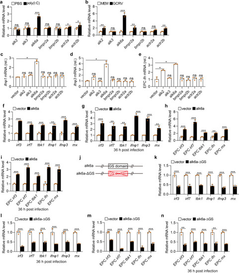
Alk6a is involved in the antiviral innate immune responses.

a, b Expression of alk2, alk3, alk6a, bmpr2a, bmpr2b, actr2a, and actr2b mRNA in ZFL cells stimulated with poly(I:C) (2 μg/ml, a) or GCRV (5 × 10⁴TCID50 per ml, b) for 48 h. ce Expression of ifnφ1 (c) and ifnφ3 (d) mRNA in ZFL cells or EPC ifn (e) in EPC cells which were transfected with 2 μg of alk2, alk3, alk6a, bmpr2a, bmpr2b, actr2a, actr2b or empty vector for 48 h. f, h Expression of irf3, irf7, tbk1, ifn (or ifnφ1 and ifnφ3), and mx mRNA after transfected with 2 μg of pcDNA3.1-alk6a or empty vector in ZFL (f) or EPC (h) cells for 48 h. g, i Expression of irf3, irf7, tbk1, ifn (or ifnφ1 and ifnφ3), and mx mRNA after transfected with 2 μg of of pcDNA3.1-alk6a or empty vector in ZFL (g) or EPC (i) cells for 24 h, followed by infection with GCRV for another 36 h. j Schematic drawing of the alk6a-ΔGS mutation that the GS domain of Alk6a was deleted. k, m Expression of irf3, irf7, tbk1, ifn (or ifnφ1 and ifnφ3), and mx mRNA after transfected with 2 μg pcDNA3.1-alk6a-ΔGS or empty vector in ZFL (k) or EPC (m) cells for 48 h. l, n Expression of irf3, irf7, tbk1, ifn (or ifnφ1 and ifnφ3), and mx mRNA after transfected with 2 μg pcDNA3.1-alk6a-ΔGS or empty vector in ZFL (l) or EPC (n) cells for 24 h, followed by infection with GCRV for another 36 h. The expression of zebrafish actb1 or EPC actin was used as an internal control for the qRT-PCR. Data were from three independent experiments and were analyzed by Student’s t-test (two-tailed) for comparison of two groups or one-way ANOVA followed by Games–Howell posthoc tests for comparison of multiple groups. All data were presented as mean ± SD (*p < 0.05, **p < 0.01, and *** p < 0.001, ns means no significant difference).

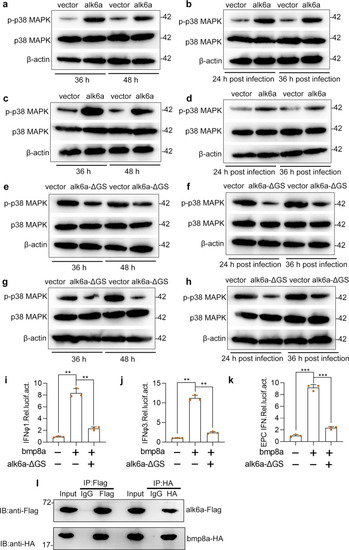
Bmp8a activates the IFN expression through Alk6a.

a, c Immunoblot analysis of phosphorylated (p-) p38 MAPK after transfected with 2 μg alk6a or empty vector in ZFL (a) or EPC (c) cells. The cells were collected at 36 or 48 h of post-transfection for Immunoblot analysis. b, d Immunoblot analysis of phosphorylated (p-) p38 MAPK after transfected with 2 μg alk6a or empty vector in ZFL (b) or EPC (d) cells for 24 h, followed by infection with GCRV (5 × 10⁴ TCID50 per ml) for another 24 h or 36 h. e, g Immunoblot analysis of phosphorylated (p-) p38 MAPK after transfected with 2 μg alk6a-ΔGS or empty vector in ZFL (e) or EPC (g) cells. The cells were collected at 36 or 48 h of post-transfection for Immunoblot analysis. f, h Immunoblot analysis of phosphorylated (p-) p38 MAPK after transfected with 2 μg alk6a or empty vector in ZFL (f) or EPC (h) cells for 24 h, followed by infection with GCRV (5 × 10⁴ TCID50 per ml) for another 24 or 36 h. ik EPC cells were cotransfected with IFN-φ1pro-luc (200 ng, i), IFN-φ3pro-luc (200 ng, j) or EPC IFNpro-luc (200 ng, k), and bmp8a (100 ng) together with or without the dominant negative plasmids alk6a-ΔGS (100 ng). At 48 h of post-transfection, the cells were collected for luciferase assays. Renilla luciferase was used as the internal control. l Co-immunoprecipitation and immunoblot analysis of EPC cells cotransfected with alk6a-Flag (1 μg) and bmp8a-HA (1 μg). Data were from three independent experiments and were analyzed by Student’s t-test (two-tailed) for comparison of two groups or one-way ANOVA followed by Games–Howell posthoc tests for comparison of multiple groups. All data were presented as mean ± SD (**p < 0.01, ***p < 0.001).

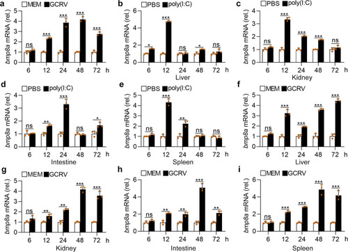
GCRV or poly(I:C) increases the expression of <italic>bmp8a</italic>.

a Expression of bmp8a mRNA in ZFL cells after infected with GCRV (5 × 10⁴ TCID50 per ml). be Expression of bmp8a mRNA in the liver (b), spleen (c), intestine (d), and kidney (e) from zebrafish injected i.p. with poly(I:C) (10 μg/fish). fi Expression of bmp8a mRNA in the liver (f), spleen (g), intestine (h), and kidney (i) from zebrafish injected i.p. with 50 µl of GCRV (108 TCID50 per ml). Zebrafish injected i.p. with PBS or MEM were used as the control. The expression of actb1 served as a control for the qRT-PCR. Data were from three independent experiments and were analyzed by Student’s t-test (two-tailed). All data were presented as mean ± SD (*p < 0.05, **p < 0.01, and ***p < 0.001, ns means no significant difference).

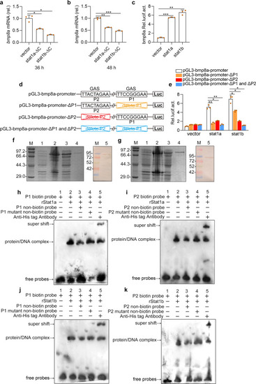
Stat1 binds the GAS sites and activates the <italic>bmp8a</italic> transcriptions upon virus stimulation.

a, b Expression of bmp8a mRNA in ZFL cells after transfected with stat1a-ΔC or stat1b-ΔC for 36 h (a) or 48 h (b). The expression of actb1 served as a control for the qRT-PCR. c Dual luciferase report assay was used to analyze the transcription abilities of Stat1a and Stat1b in activation of bmp8a in EPC cells. pGl3-bmp8a (200 ng) was transfected into EPC cells together with stat1a (200 ng), stat1b (200 ng), or empty vector (200 ng). After 48 h, the transfected cells were collected for luciferase assays. d Schematic drawing of wild-type and GAS motif mutation Luc-report plasmids. e The 200 ng of pGL3-bmp8a-promoter, pGL3-bmp8a-promoter-ΔP1, pGL3-bmp8a-promoter-ΔP2 or pGL3-bmp8a-promoter-ΔP1 and ΔP2 was transfected into EPC cells along with stat1a (200 ng), stat1b (200 ng) or empty vector (200 ng), respectively. After 48 h, the transfected cells were collected for luciferase assays. Renilla luciferase was used as the internal control. f, g SDS-PAGE and Western-blotting analysis of rStat1a (f) and rStat1b (g). Lane M: protein molecular standard; Lane 1: negative control for IPTG induced E. coli (without rStat1a or rStat1b); Lane 2: induced rStat1a or rStat1b (the whole cell lysate); Lane 3: induced rStat1a or rStat1b (the supernatant); Lane 4: purified rStat1a or rStat1b; Lane 5: western blot analysis of the sample in Lane 4. hk EMSA was performed to validate the interaction of rStat1a or rStat1b with the GAS motif (P1 or P2) in the bmp8a promoter region. Lane 1: negative group; Lane 2: positive group; Lane 3: an excess unlabeled competitor probe; Lane 4: an excess unlabeled competitor probe containing a mutated runt binding site; Lane 5: Super-shift assays were performed by adding antibody against His tag. Data were from three independent experiments and were analyzed by Student’s t-test (two-tailed) for comparison of two groups or one-way ANOVA followed by Games–Howell posthoc tests for comparison of multiple groups. All data were presented as mean ± SD (*p < 0.05, **p < 0.01, and ***p < 0.001).

Schematic illustration of Bmp8a serving as positive regulator in the antiviral immune.

Upon virus infection, the transcriptions of bmp8a are activated through the Jak-Stat1 pathway. The Bmp8a binds to BMP type I receptor Alk6a, promoting phosphorylation of Tbk1 and Irf3 to induce the expression of Ifn through p38 MAPK pathway.

Acknowledgments
This image is the copyrighted work of the attributed author or publisher, and ZFIN has permission only to display this image to its users. Additional permissions should be obtained from the applicable author or publisher of the image. Full text @ Commun Biol