
- Title
-
The Redox Activity of Protein Disulfide Isomerase Inhibits ALS Phenotypes in Cellular and Zebrafish Models
- Authors
- Parakh, S., Shadfar, S., Perri, E.R., Ragagnin, A.M.G., Piattoni, C.V., Fogolín, M.B., Yuan, K.C., Shahheydari, H., Don, E.K., Thomas, C.J., Hong, Y., Comini, M.A., Laird, A.S., Spencer, D.M., Atkin, J.D.
- Source
- Full text @ iScience

The Oxidoreductase Activity of PDI Is Protective against Inclusion Formation and Protein Unfolding in Mutant TDP-43 Expressing Cells. (A) Immunoblotting was performed to confirm that similar transfection efficiencies were present and that co-expression of PDI-WT or PDI-QUAD did not alter the expression of TDP-43 EGFP. An anti-TDP-43 antibody was used to detect the presence of wild-type TDP-43 (TDP-WT) or mutant TDP-43M337V (TDP-M337V), in cells co-expressing either empty vector pcDNA3.1 or PDI-V5 (WT or QUAD), β-actin was used as a loading control. (B) Immunofluorescence detection of EGFP in cells expressing EGFP-tagged TDP-WT (row 1), TDP-M337V with empty vector alone (row 2) or co-expressing PDI-WT or PDI-QUAD, or administered with BMC (rows 3, 4, 5). (C) Significantly fewer cells formed inclusions when PDI-WT was co-expressed with TDP-M337V or treated with BMC (∗∗p < 0.01). Significant differences were observed between TDP-M337V cells co-expressing PDI-WT or PDI-QUAD, and TDP-M337V cells co-expressing PDI-QUAD or BMC (∗∗p < 0.01). (D) Neurons expressing EGFP only (row 1), TDP-WT (row 2), TDP-M337V alone (row 3), or co-expressing PDI-WT or PDI-QUAD, or BMC-treated (rows 4, 5, 6). (E) Significantly fewer cells formed inclusions when PDI-WT was co-expressed with TDP-M337V (∗∗p < 0.01) and treated with BMC (∗p < 0.05). Significant differences were observed between TDP-M337V cells co-expressing PDI-WT or PDI-QUAD (∗p < 0.05). (F) TPE-MI fluorescence in Neuro-2a cells expressing TDP-WT (row 1), TDP-M337V with empty vector alone (row 2), or co-expressing PDI-WT or PDI-QUAD, or treated with BMC (rows 3, 4, 5), arrows represent TPE-MI fluorescence. (G) Significantly fewer cells displayed TPE-MI fluorescence (representing the cellular load of unfolded proteins, blue) when PDI-WT was co-expressed with TDP-M337V or cells were treated with BMC (∗∗p < 0.01) compared to controls. Scale bars: 8 μm in (B), 5 μm in (D), 12 μm in (F). |

The Oxidoreductase Activity of PDI Is Protective against TDP-43 Mislocalization to the Cytoplasm (A) Neuro-2a cells expressing EGFP-tagged TDP-WT (row 1), TDP-M337V with empty vector (row 2), co-expressing PDI-WT or PDI-QUAD, or treated with BMC (rows 3, 4, 5), arrows repesent mislocalised TDP-43. (B) Expression of PDI-WT (∗∗p < 0.01) or administration of BMC (∗p < 0.05) to mutant TDP-M337V-expressing cells significantly reduced the proportion of cells displaying cytoplasmic TDP-43. A significant difference in cells expressing cytoplasmic TDP-43 was observed between PDI-WT and PDI-QUAD (∗∗p < 0.01), and between PDI-QUAD and BMC (∗p < 0.05). (C) Primary neurons expressing TDP-WT (row 1), TDP-M337V with empty vector (row 2), co-expressing PDI-WT or PDI-QUAD, or treated with BMC (rows 3, 4, 5), arrows repesent mislocalised TDP-43. (D) Over-expression of PDI-WT (∗∗p < 0.01) or BMC treatment (∗p < 0.05) in mutant TDP-M337V-expressing cells significantly reduced the proportion of cells displaying cytoplasmic TDP-43. There was also a significant difference between TDP-M337V cells co-expressing PDI-WT and those co-expressing PDI-QUAD (∗p < 0.05). (E) Neuro-2a cells expressing mCherry-tagged TDP-WT (row 1), TDP-Q331K (row 2), or co-expressing PDI-WT or PDI-QUAD, or treated with BMC (rows 3, 4, 5), arrows repesent mislocalised TDP-43 (F) Expression of PDI-WT or treatment with BMC (∗∗∗p < 0.001) in mutant TDP-Q331K-expressing cells significantly reduced the proportion of cells displaying cytoplasmic TDP-43, compared with cells expressing empty vector only. A significant difference was observed between PDI-WT and PDI-QUAD, and between PDI-QUAD and BMC treated cells (∗p < 0.05). Scale bars: 10 μm in (A) and (E), 5 μm in (C). |
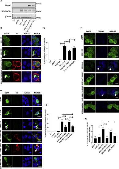
The Oxidoreductase Activity of PDI Is Protective against Inclusion Formation and Protein Unfolding in Mutant SOD1 expressing cells Immunoblotting was performed to confirm that similar transfection efficiencies were present and that co-expression of PDI-WT or PDI-QUAD did not alter the expression of SOD1. An anti-GFP antibody was used to detect SOD1-WT or mutant SOD1A4V (SOD1-A4V), in cells co-expressing empty vector pcDNA3.1 or PDI-V5 (WT or QUAD). β-actin was used as a loading control (bottom panel). (B) Immunofluorescence detection of EGFP in cells expressing SOD1-WT (row 1) or SOD1-A4V (inclusions represented by white arrows, row 2), co-expressed with PDI-WT or PDI-QUAD (rows 3, 4). (C) Significantly fewer cells formed inclusions when PDI-WT was co-expressed with SOD1-A4V (∗∗∗p < 0.001), and significant difference was observed between PDI-WT and PDI-QUAD expressing cells (∗p < 0.05). (D) Immunofluorescence detection of EGFP-positive inclusions present in mouse primary neurons co-expressing EGFP only (row 1), SOD1-WT (row 2) or SOD1-A4V (row 3), with PDI-WT or PDI-QUAD, or treated with BMC (rows 4, 5, 6). (E) Significantly fewer neurons formed inclusions when PDI-WT was co-expressed with SOD1-A4V (∗∗p < 0.01) or treated with BMC (∗p < 0.05). A significant difference was observed between SOD1-WT and mutant SOD1-A4V (∗∗∗∗p<0.0001) cells. n = 35, ANOVA followed by Tukey's post hoc test. (F) TPE-MI fluorescence in Neuro-2a cells expressing pEGFP (row 1), SOD1-WT (row 2), or SOD1-A4V cells (row 3), co-expressing PDI-WT or PDI-QUAD, or treated with BMC (rows 4, 5, 6). (G) Significantly fewer cells displayed TPE-MI fluorescence when PDI-WT was co-expressed with SOD1-A4V or treated with BMC (∗∗p < 0.01 and ∗p < 0.05). Scale bars: 10 μm in (B), 10 μm in (D), 15 μm in (F). |
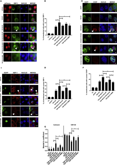
The Oxidoreductase Activity of PDI Is Protective against ER Stress and Inhibition of ER-Golgi Transport Induced by Mutant TDP-43 (A) Detection of nuclear immunoreactivity to XBP-1 in cells expressing mCherry-tagged TDP-43. Cells expressing mCherry (row 1), TDP-WT (row 2), or mutant TDP-Q331K (row 3), co-expressing PDI-WT or PDI-QUAD, or treated with BMC (rows 4, 5, 6), arrows representing XBP-1 activation. (B) The proportion of cells expressing nuclear XBP-1 decreased when PDI-WT was co-expressed or treated with BMC (∗p < 0.05), unlike PDI-QUAD. (C) Immunofluorescence detection of nuclear immunoreactivity to XBP-1 in EGFP-tagged TDP-43 cells. Cells expressing TDP-WT (row 1), or TDP-M337V (row 2), co-expressing PDI-WT or PDI-QUAD, or treatment with BMC (rows 3, 4, 5), arrows represent XBP-1 activation. (D) The proportion of cells expressing nuclear XBP-1 decreased when PDI-WT was co-expressed, or BMC was administered to TDP-M337V cells (∗∗p < 0.01). More cells with nuclear XBP-1 were found in populations expressing PDI-QUAD compared with PDI-WT, and PDI-QUAD compared with BMC treatment (∗p < 0.05). (E) Detection of nuclear immunoreactivity to CHOP in EGFP-tagged TDP-43 cells. Cells expressing pEGFP (row 1), TDP-WT (row 2), TDP-M337V (row 3), co-expressing PDI-WT or PDI-QUAD, or treated with BMC (rows 4, 5, 6), arrows represent CHOP activation. (F) The proportion of cells expressing nuclear CHOP was decreased when PDI-WT was co-expressed or BMC was administered to TDP-M337V cells (∗∗p < 0.01). There was a significant difference between TDP-M337V cells co-expressing PDI-WT and PDI-QUAD (∗∗p < 0.01), and TDP-M337V cells co-expressing PDI-QUAD and treated with BMC (∗∗p < 0.01). (G) PDI's oxidoreductase activity rescues inhibition of ER-Golgi transport induced by mutant TDP-43. Quantification of the degree of co-localization of VSVGts045 with the ER and Golgi compartments using Mander's coefficient following immunocytochemistry for calnexin and GM130. Data are presented as mean ± SEM, n = 20. A significant difference was observed (∗p < 0.05) in the co-localization between VSVGts045 and the ER (calnexin) between cells expressing TDP-Q331K with empty vector and those expressing PDI-WT, and also with (∗p < 0.05) BMC-treatment . A significant difference was also observed (∗p < 0.05) in co-localization between VSVGts045 and the Golgi (GM130) between cells expressing TDP-Q331K and PDI-WT and BMC-treated cells. Scale bars: 10 μm in (A) and (C), 4 μm in (E). |
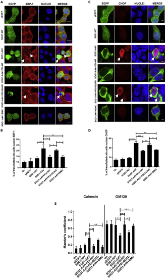
The Oxidoreductase Activity of PDI Is Protective against ER Stress and Inhibition of ER-Golgi Transport Induced by Mutant SOD1 (A) Detection of nuclear immunoreactivity to XBP-1 (second column) in EGFP (row 1), SOD1-WT (row 2) or SOD1-A4V (row 3) cells, co-expressing either PDI-WT or PDI-QUAD, or treated with BMC (rows 4, 5, 6), arrows represent XBP-1 activation. (B) Fewer cells expressing nuclear XBP-1 were present when PDI-WT was co-expressed or cells were treated with BMC (∗∗p < 0.01). There was a significant difference between SOD1-WT and SOD1-A4V cells (∗∗∗p<0.001). Similarly, a significant difference was observed between SOD1-A4V cells co-expressing PDI-WT or PDI-QUAD, and SOD1-A4V cells co-expressing PDI-QUAD or treated with BMC (∗p < 0.05). (C) Immunofluorescence detection of nuclear immunoreactivity to CHOP (second column) in cells expressing EGFP (row 1), SOD1-WT only (row 2), or SOD1-A4V (row 3) with PDI-WT or PDI-QUAD, or treated with BMC (rows 4, 5, 6), arrows represent CHOP activation. (D) The proportion of cells expressing nuclear CHOP was significantly decreased when PDI-WT was co-expressed with SOD1-A4V (∗∗∗p < 0.001) or treated with BMC (∗∗p < 0.01). There was significant difference between SOD1-WT and SOD1-A4V cells (∗∗∗∗p<0.0001). Significant differences were also detected between SOD1-A4V cells co-expressing PDI-WT and PDI-QUAD (∗∗p < 0.01), and SOD1-A4V cells co-expressing PDI-QUAD or treated with BMC (∗p < 0.05). (E) PDI's oxidoreductase activity rescues inhibition of ER-Golgi transport induced by mutant SOD1. Quantification of the degree of co-localization of VSVGts045 with the ER and Golgi compartments using Mander's coefficient following immunocytochemistry for calnexin and GM130. Data are presented as mean ± SEM, n = 20. More co-localization between VSVGts045and the ER (calnexin) was observed (∗∗p < 0.01) in SOD1-A4V cells compared with those co-expressing PDI-WT or treated with BMC. More SOD1-A4V and PDI-WT co-expressing cells, or those treated with BMC, displayed co-localization between VSVGts045 and the Golgi (GM130) (∗∗∗p < 0.001). Similarly, there was a significant difference in Golgi localisation between cells expressing PDI-WT and PDI-QUAD (∗p < 0.05). Scale bars: 10 μm in (A), 4 μm in (C). |
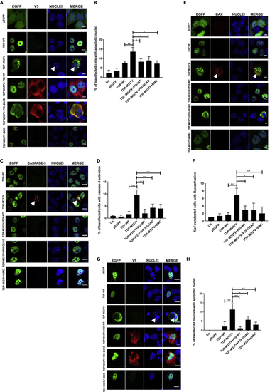
The Oxidoreductase and Chaperone Activities of PDI Are Protective against Mutant TDP-43 Induced Cell Death (A) Cells expressing EGFP (row 1), TDP-WT (row 2) or TDP-M337V (row 3), co-expressing PDI-WT or PDI-QUAD or BMC treated (rows 4, 5, 6), arrows represent apoptotic nuclei. (B) Over-expression of PDI-WT, PDI-QUAD, or BMC treatment with TDP-M337V resulted in significantly fewer cells with apoptotic nuclei compared with cells transfected with empty vector (∗p < 0.05). A significant difference was observed between TDP-WT and TDP-M337V expressing cells (∗∗p<0.01). (C) Immunocytochemistry using activated caspase-3 antibodies (red). Cells expressing TDP-WT (row 1) or TDP-M337V (row 2, arrows representing caspase-3 activation), co-expressing PDI-WT or PDI-QUAD, or treated with BMC (rows 3, 4, 5). (D) Over-expression of PDI-WT (∗∗∗p < 0.001), PDI-QUAD or treatment with BMC (∗∗p < 0.01), significantly decreased the proportion of cells with activated caspase-3. (E) Immunocytochemistry using anti-activated Bax antibodies (red). Cells expressing pEGFP (row 1), TDP-WT (row 2), or TDP-M337V (row 3, arrows representing Bax activation), co-expressing PDI-WT or PDI-QUAD, or treated with BMC (rows 4, 5, 6). (F) Over-expression of either PDI-WT or PDI-QUAD (∗p < 0.05), or treatment with BMC (∗∗p < 0.01), significantly decreased the proportion of cells with activated Bax compared with cells expressing empty vector. (G) Primary neurons expressing EGFP (row 1), TDP-WT (row 2) or TDP-M337V (row 3), co-expressing PDI-WT, PDI-QUAD, or treated with BMC (rows 4, 5, 6), arrows represent apoptotic nuclei. (H) Mutant TDP-M337V expression induced apoptosis (∗∗∗p < 0.001); however, over-expression of PDI-WT (∗∗∗p < 0.001), PDI-QUAD (∗p < 0.05) or administration of BMC (∗∗p < 0.01) resulted in significantly fewer neurons undergoing apoptosis. Scale bars: 8 μm in (A), (G), 10 μm in (C), 4 μm in (E). |
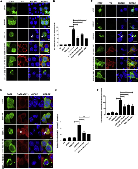
The Oxidoreductase and Chaperone Activities of PDI Are Protective against Mutant SOD1 Induced Cell Death (A) Neuro-2a cells expressing EGFP (row 1), SOD1-WT (row 2) or SOD1-A4V (row 3, condensed nuclei represented by white arrows), SOD1-A4V co-expressing PDI-WT or PDI-QUAD, or BMC treatment (rows 4, 5, 6). (B) Over-expression of PDI-WT (∗∗∗∗p < 0.0001), PDI-QUAD (∗∗p < 0.01) or BMC (∗∗∗∗p < 0.0001) with SOD1-A4V resulted in significantly fewer condensed apoptotic cells. A significant difference was observed between SOD1-WT and SOD1-A4V expressing cells (∗∗∗∗p<0.0001). (C) Immunocytochemistry using activated caspase-3 antibodies (red), white arrow represents caspase-3 activation. Cells expressing EGFP (row 1), SOD1-WT (row 2), SOD1-A4V (row 3), or co-expressing SOD1-A4V and PDI-WT or PDI-QUAD (rows 4, 5). (D) Over-expression of both PDI-WT (∗∗p < 0.01) and PDI-QUAD (∗∗∗p < 0.001) with SOD1-A4V significantly decreased the proportion of cells with activated caspase-3. (E) Primary neurons expressing pEGFP (row 1), SOD1-WT (row 2), or SOD1-A4V (row 3), co-expressing PDI-WT or PDI-QUAD, or treatment with BMC (rows 4, 5, 6). (F) Co-expression of PDI-WT (∗∗∗p < 0.001) or PDI-QUAD (∗∗p < 0.01) or treatment with BMC (∗∗∗p < 0.001) in SOD1-A4V expressing cells resulted in significantly fewer cells with apoptotic nuclei, identified by the presence of activated caspase-3. A significant difference was observed between SOD1-WT and SOD1-A4V (∗∗∗∗p<0.0001). Scale bars: 4 μm in (A) and (C), 10 μm in (E). |

ALS-Linked PDI Mutants (D292N and R300H) Are Not Protective against Mutant TDP-43 and Mutant SOD1 (A) Cells expressing TDP-M337V with empty vector alone (row 1) or co-expressing PDI-WT, PDI-D292N or PDI-R300H (rows 2, 3, 4). (B) Significantly fewer cells formed inclusions when PDI-WT was co-expressed with TDP-M337V (∗p < 0.05) compared with PDI-D292N and PDI-R300H (∗p < 0.05). (C) Cells expressing EGFP-tagged TDP-M337V (row 1), or co-expressing PDI-WT, PDI-D292N or PDI-R300H (rows 2, 3, 4). (D) Co-expression of PDI-WT (∗p < 0.05) with mutant TDP-M337V significantly reduced the proportion of cells with cytoplasmic TDP-43 expression compared with those co-expressing PDI-D292N or PDI-R300H (∗p < 0.05). (E) Cells expressing SOD1-A4V (row 1) or co-expressing PDI-WT, PDI-D292N or PDI-R300H (rows 2, 3, 4). (F) Significantly fewer cells with inclusions were observed when PDI-WT was co-expressed with SOD1-A4V (∗p < 0.05) compared with either PDI D292N (∗∗p < 0.01) or PDI R300H (∗p < 0.01). (G) Neuro-2a cells expressing SOD1-A4V (row 1) or co-expressing PDI-WT, PDI-D292N or PDI-R300H (rows 2, 3, 4). (H) Over-expression of PDI-WT (∗∗p < 0.01) with SOD1-A4V resulted in significantly fewer cells with apoptotic nuclei compared with those expressing empty vector and those co-expressing PDI-D292N or PDI-R300H (∗p < 0.05). Scale bars: 6 μm in (A), (C), (E), and (G). |