FIGURE SUMMARY
Title

Exploiting Zebrafish Xenografts for Testing the in vivo Antitumorigenic Activity of Microcin E492 Against Human Colorectal Cancer Cells

Authors
Varas, M.A., Muñoz-Montecinos, C., Kallens, V., Simon, V., Allende, M.L., Marcoleta, A.E., Lagos, R.
Source
Full text @ Front Microbiol

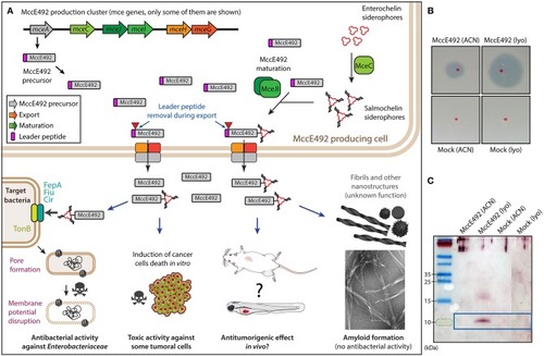
(A) Main features of microcin E492 peptide and its production by bacterial cells, through the expression of the mce genes from the MccE492 gene cluster. The mceA gene encodes the microcin precursor that includes a leader peptide that is removed during the export by ABC exporters encoded by mceHG. During maturation, salmochelin (a glycosylated enterochelin derivative synthetized by MceC) is attached to MccE492 C-terminus by MceIJ. Mature MccE492 is recognized and internalized by catechol siderophore receptors of the target bacterial cell, and once in the periplasm it inserts into the cytoplasmic membrane in a TonB-dependent process forming pores that lead to a toxic effect through the disruption of the membrane potential of the cell. Additionally, under certain conditions, MccE492 aggregates into amyloid fibers and rod-shaped nanostructures, leading to the loss of its antibacterial activity. MccE492 also has toxic activity in vitro against some tumor cell lines, although its in vivo antitumoral effect has not been validated in animal models. For simplicity, several genes from the MccE492 gene cluster are not included, among them the immunity gene mceB that is encoded in an operon with the structural gene mceA. (B) Antibacterial activity assay of a representative MccE492 preparation (either the ACN fraction or after lyophilization) assessed through the detection of growth inhibition halos over sensitive bacterial lawns. The red dots correspond to the zone of the lawn where the drops of MccE492 or the mock preparation were placed. (C) SDS-PAGE and immunoblot of a representative MccE492 preparation (either the ACN fraction or after lyophilization), revealed with a monoclonal anti-McccE492 antibody. The last lane of the immunoblot image (showing mock purification) was spliced together with the first 3 lanes, to avoid showing blank (non-loaded) lanes.

Microcin E492 induces cytotoxicity over human colorectal cancer cell lines in vitro. HT29 (A) and SW620 (B) cells were incubated during 24 h with MccE492, a mock purification, or PBS, and the cell viability was determined through flow cytometry using the Sytox Green Dead Cell stain (Invitrogen) and liquid counting beads. Colored areas denote the three populations distinguished (P1-P3), and the numbers under each population label indicate the percentage from the total population. Each plot shows a total of 10,000 cells.

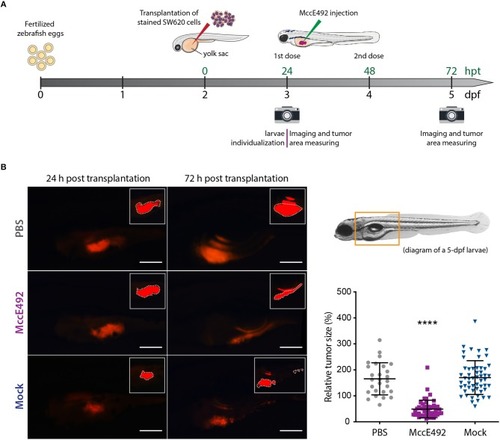
Microcin E492 intratumoral injection reduces the tumor size in zebrafish larvae SW620 xenograft model. (A) Schematic representation of the experimental setup for the development of zebrafish xenografts using SW620 human colorectal cancer cells to test the MccE492 antitumoral effect. (B) Assessment of the tumor cell mass size through live-cell imaging in zebrafish xenografts upon injection of MccE492, a mock purification, or PBS. Representative images of each condition are shown in the left side. The photographed area is delineated in the 5-dpf larva scheme shown. The insets show the saturated fluorescence area used in the estimation of tumors size (only the zones inside the white dashed lines were considered, as the excluded red zones correspond to reflections of the fluorescence signal in the swim bladder). The relative tumor size, corresponding to the percent change in tumor size comparing the same individual at 24 and 72 hpt, was determined and plotted for a total cohort of 50 individuals per condition. Error bars correspond to the standard deviation. ****P < 0.0001 (two-way ANOVA with a Dunnet's post-test).

PHENOTYPE:
Fish:
Condition:
Observed In:
Stage: Day 5
Acknowledgments
This image is the copyrighted work of the attributed author or publisher, and ZFIN has permission only to display this image to its users. Additional permissions should be obtained from the applicable author or publisher of the image. Full text @ Front Microbiol