
- Title
-
Temporal control of Wnt signaling is required for habenular neuron diversity and brain asymmetry
- Authors
- Guglielmi, L., Bühler, A., Moro, E., Argenton, F., Poggi, L., Carl, M.
- Source
- Full text @ Development
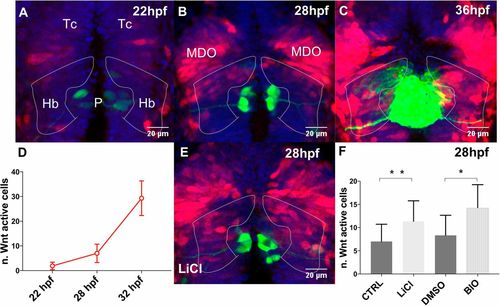
Wnt signaling activity in and around the developing habenulae. (A-C,E) Projections of confocal z-stacks, dorsal views, anterior is towards the top focused onto the diencephalon of tg(7xtcf-Xla.Siam:nls-mCherry); tg(flh:GFP); tg(foxD3:GFP) transgenic embryos at stages indicated. Nuclei are blue, Wnt active cells are red and the pineal complex is green. Dotted lines highlight the region of the developing habenulae. (D) Graph shows the number of Wnt-active habenular precursors, which are (F) increased, when Wnt signaling is activated by drug treatments, as indicated. Hb, habenulae; MDO, mid-diencephalic organizer; P, pineal; Tc, telencephalon. EXPRESSION / LABELING:
PHENOTYPE:
|

Premature intrinsic activation of Wnt signaling delays habenular neuron differentiation. (A-B′) Projections of confocal z-stacks, dorsal views, anterior is towards the top focused onto the diencephalon of (A,A′) Et(-1.0otpa:mmGFP) and (B,B′) tg(flh:GFP); tg(foxD3:GFP) transgenic embryos. Nuclei are DAPI labeled (purple). (A,A′,C) Transient Wnt signaling activation causes a specific loss of GFP-expressing habenular neurons at 48 hpf. (B,B′,D,E) The number of HuC/D-positive differentiating habenular neurons is reduced at 36 hpf; their left-right asymmetric development remains unchanged. CTRL, control; Hb, habenulae; P, pineal; Tec, optic tectum; Th, thalamus. |
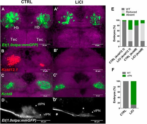
Premature activation of Wnt signaling causes a reduction of dHbl neurons. (A-D′) Projections of confocal z-stacks. (A-C′) Dorsal views, anterior is towards top focused onto the diencephalon of (A-D′) embryos at 3 dpf. Nuclei are DAPI labeled (purple). (A,A′) LiCl treatment causes delayed habenular neuron differentiation. (B-C′,E) Markers for (B,B′) dHbl and (C,C′) dHbm neurons are reduced as represented in E. (D,D′) Lateral views of IPN innervation by habenular efferent axons, anterior is leftwards. Treated embryos show vIPN innervation, as represented in F. d, dorsal; Hb, habenula, IPN, interpeduncular nucleus; Tec, optic tectum; v, ventral. EXPRESSION / LABELING:
PHENOTYPE:
|

Wif1 controls dHb neuron differentiation and is regulated by Wnt signaling. (A-C′) Projections of confocal z-stacks. (A-B′) Dorsal views, anterior is towards the top focused onto the diencephalon of embryos at 3 dpf. Nuclei are DAPI labeled (purple). (A-B′,D,E) Wif1 hypomorphic embryos exhibit delayed habenular neuron differentiation (asterisks in A′,B′). (C,C′) Lateral views focused on the IPN of Et(-1.0otpa:mmGFP) embryos with anterior towards the left. (C,C′,F) Wif1 knockdown embryos show predominant vIPN innervation. Asterisk highlights the missing dIPN innervation. (G-I) Lateral views focused on the diencephalon of 25 hpf (G) untreated and (H,I) treated embryos, labeled for Wif1 expression. Scale bar: 60 µm. Labeling procedures were conducted in parallel for comparison. d, dorsal; Hb, habenula, IPN, interpeduncular nucleus; P, pineal; v, ventral. EXPRESSION / LABELING:
PHENOTYPE:
|