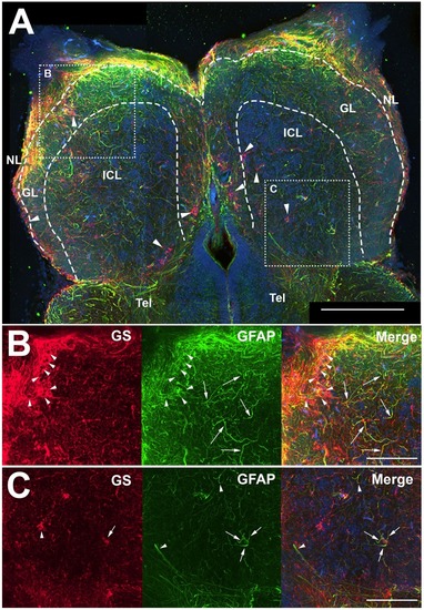
Z-stack images of anti-glial fibrillary acidic protein (GFAP) and anti-glutamine synthetase (GS) immunoreactivity in the olfactory bulb of uninjured adult zebrafish. (A) There was a high degree of labeling for both antibodies in the nerve layer (NL) and glomerular layer (GL) of the olfactory bulb that varied in co-labeling. Anti-GS (red) labeling did not reveal clear structures with the exception of ring-like labeling seen in all layers of the bulb (arrowheads). There were numerous, distinct anti-GFAP (green) labeled processes in the GL and internal cell layer (ICL) that varied in size and length but had no obvious organization. Tel, Telencephalon. (B) Higher magnification of the rostral bulb region shown in (A) confirmed that ring-like structures (arrowheads) were labeled mostly with anti-GS and distinct processes (arrows) were labeled predominantly with anti-GFAP. (C) Higher magnification of the caudal bulb region revealed less labeling with both antibodies in this region, but anti-GS positive ring-like structures and anti-GFAP positive processes were apparent. Scale bar = 100 μm (A) or 20 μm (B,C).

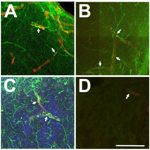
Relationship between anti-GFAP labeling and capillaries viewed in Z-stack images. (A–C) Anti-GFAP processes (green) often appeared to terminate onto capillaries (arrows), identified by their tubular appearance and red auto-fluorescence. (D) Antibody controls where the primary antibody was eliminated show low levels of green background staining and red auto-fluorescent capillaries (arrow). Scale bar = 20 μm for all.

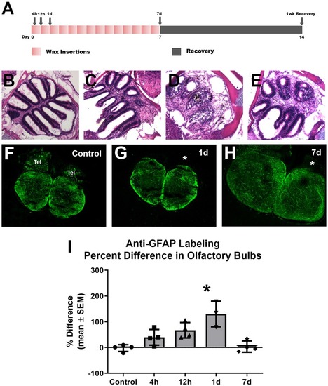
Gross analysis with Z-stack images of anti-GFAP labeling in the olfactory bulb following repetitive peripheral damage. (A) Illustration of the treatment paradigm shows that fish receiving repeated wax plug insertions were analyzed at various survival times during wax treatment and after a recovery period. Images of olfactory organs show typical rosette morphology in control fish (B), significant damage at 1 day (C) and 7 days (D) of wax plug insertions, and return to control morphology with 1 week of recovery (E) after removal of the plug. (F) Untreated control fish had typical anti-GFAP labeling in all layers of both olfactory bulbs. Tel = Telencephalon, n = 4. (G) After 1 day of repetitive peripheral damage to the olfactory organ, the affected bulb (asterisk) appeared to have slightly more anti-GFAP labeling than the internal control bulb, n = 3. (H) By 7 days, there was no noticeable difference in labeling between bulbs, and anti-GFAP labeling in the affected bulb (asterisk) appeared at control levels, n = 4. Scale bar = 100 μm for all.(I) A comparison of anti-GFAP labeling between bulbs showed a significant difference at 1 day of repetitive peripheral damage when compared to controls but no difference at 7 days of damage. *p < 0.05.

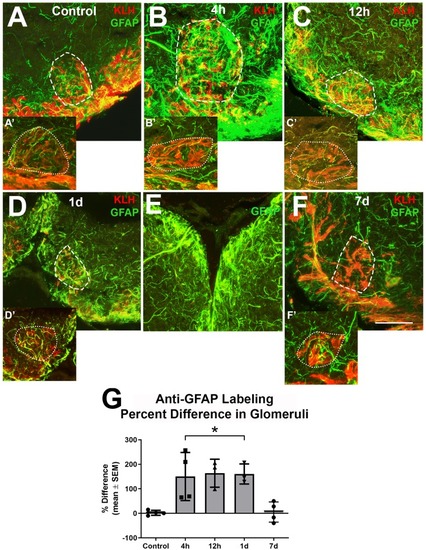
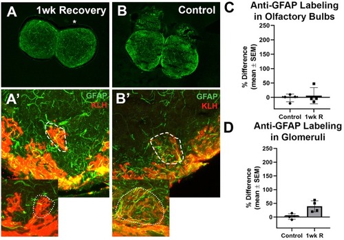
Higher magnification analysis of Z-stacks of anti-GFAP (green) and anti-keyhole limpet hemocyanin (KLH; red) labeling during repetitive damage to the olfactory organ. (A) Anti-KLH allowed identification of a ventral medial glomerulus (dashed outline). Anti-GFAP labeled processes within the glomerulus in the right olfactory bulb (dashed outline) varied in size and length and had no clear organization in untreated control bulbs. (A’) A glomerulus (dotted outline) in the same region of the left olfactory bulb of the same fish displayed similar morphology of anti-GFAP labeled processes, n = 4. (B) After 4 h of damage to the olfactory organ, anti-GFAP labeled processes appeared to be more numerous and thicker within the glomerulus (dashed outline) of the affected bulb when compared to the comparable glomerulus (dotted outline) of the internal control bulb (B’), n = 4. (C) By 12 h after damage to the periphery, there were numerous anti-GFAP labeled processes associated with the glomerulus (dashed outline) in the affected bulb that varied in length and thickness, while the glomerulus (dotted outline) in the internal control bulb (C’) retained typical labeling, n = 3. (D) At 1 day after repeated damage to the olfactory organ, affected glomeruli (dashed outline) still appeared to have thicker and more numerous anti-GFAP labeled processes when compared to glomeruli (dotted outline) in the internal control bulb (D’), n = 3. (E) Side by side comparison of both bulbs at 1 day showed that the left, internal control bulb retained control levels of anti-GFAP labeling, while the right, treated bulb showed more and thicker labeled processes. (F) By 7 days after repetitive peripheral damage, anti-GFAP labeled processes in glomeruli (dashed outline) in the affected bulb appeared to have similar morphology to anti-GFAP labeled processes in glomeruli (dotted outline) of the internal control bulb (F’), n = 4. Scale bar = 20 μm for all. (G) The percent difference of anti-GFAP optical density (OD) measurements between treated and untreated glomeruli was significantly different at 4 h, 12 h, and 1 day of repetitive peripheral damage but was no longer significantly different after 7 days of repeated damage. *p < 0.05.

The recovery of the olfactory bulb does not involve evidence of a glial scar. (A) After the olfactory system was allowed 1 week to recover from 7 days of repetitive peripheral damage, there was no apparent difference in the overall amount of anti-GFAP (green) and anti-KLH (red) labeling in the affected bulb (asterisk) when compared to the internal control bulb (n = 4). There was also no noticeable difference in the overall amount of anti-GFAP labeling compared to untreated control fish (B). (A’) Higher magnification Z-stack images revealed that anti-GFAP labeling (green) within anti-KLH (red) labeled glomeruli (dotted line) in the affected bulb after 1 week of recovery also appeared to be similar to that of glomeruli in the internal control bulb (dotted outline in inset) and in untreated control bulbs (B’) on the right (dashed outline) and left (dotted outline in inset) sides. There was no significant difference in the percent difference of OD of anti-GFAP labeling in the olfactory bulb (C) or ventral medial glomeruli (D) of zebrafish that were allowed 1 week of recovery from 7 days of repetitive peripheral damage.

Acknowledgments
This image is the copyrighted work of the attributed author or publisher, and ZFIN has permission only to display this image to its users. Additional permissions should be obtained from the applicable author or publisher of the image. Full text @ Front. Neuroanat.