FIGURE SUMMARY
Title

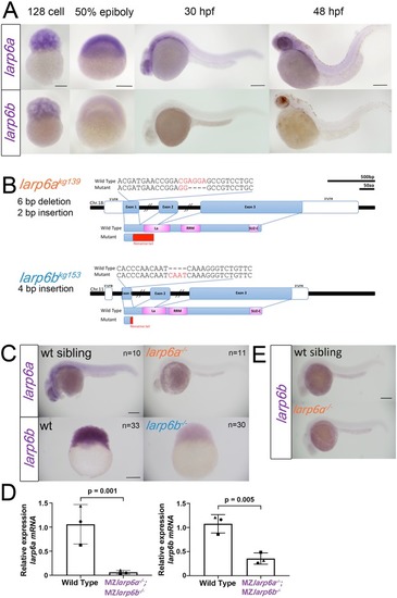
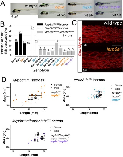
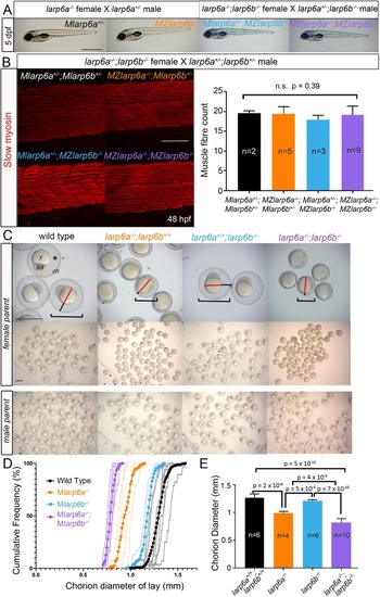
Maternal Larp6 controls oocyte development, chorion formation and elevation

Authors
Hau, H.T.A., Ogundele, O., Hibbert, A.H., Monfries, C.A.L., Exelby, K., Wood, N.J., Nevarez-Mejia, J., Carbajal, M.A., Fleck, R.A., Dermit, M., Mardakheh, F.K., Williams-Ward, V.C., Pipalia, T.G., Conte, M.R., Hughes, S.M.
Source
Full text @ Development

Genome editing generates likely null alleles of zebrafish larp6a and larp6b. (A) In situ RNA hybridisation for larp6a and larp6b at the indicated stages. (B) Schematic of larp6a and larp6b genes and proteins showing the position of kg139 and kg153 mutant alleles. The larp6akg139 frameshift mutation produces a truncated protein with the first 29 amino acids of Larp6a followed by a 72 amino acid tail lacking both La motif and RRM domains. The larp6bkg153 frameshift allele truncates Larp6b at amino acid 17 with an eight amino acid tail terminating in coding exon 1. There is a lack of in-frame ATG codons near the termination site. (C) In situ RNA hybridization for larp6a mRNA on genotyped larp6akg139 mutant and wild-type siblings from a larp6akg139/+ incross reveals nonsense-mediated mRNA decay (NMD) of mutant larp6akg139 mRNA at 24 hpf. Eleven out of 47 low expressors were shown to be mutant and 10/47 normal expressors were wild type upon sequence genotyping. As larp6b mRNA is primarily maternally expressed, NMD was analysed at the 256-cell stage by larp6b mRNA in situ RNA hybridization on lays from incrosses of larp6bkg153 F3 mutants or their wild-type siblings (see Table S1). The results shown were observed in 33/33 embryos from a wild-type female and 30/30 from a mutant female. Genotypes of mutant embryos shown were confirmed by sequencing. (D) QRT-PCR on RNA from 1k stage embryos from wild-type or larp6akg139;larp6bkg153 double mutant incrosses confirmed reduction of each mutant mRNA. Symbols indicate results from three individual RNA preparations from three separate pairs of lays. (E) Lack of larp6b mRNA upregulation in larp6akg139 mutants. All images are lateral views with anterior to the left and dorsal upwards, except 128 cell and 50% epiboly, which are animal upward. Scale bars: 200 µm.

Zygotic Larp6 mutants appear wild type. (A) Bright-field images of 5 dpf larvae from wild-type and incrosses of single and double heterozygous carriers. Fish are shown anterior towards the left and dorsal upwards with genotyped mutants above their respective wild-type siblings (wt sib). Inflated swim bladders show that larvae have swimming and swallowing capacity. (B) Adults derived from incrosses of larp6a+/ (n=52), larp6b+/ (n=62) and larp6a+/−;larp6b+/− (n=61) fish were co-reared, then genotyped at 5 mpf showing the expected Mendelian ratios. Fish numbers are above each bar. (C) Slow myosin immunofluorescence of 24 hpf embryos. Images of wild type and mutant centred on somite 17/18 reveal no differences in slow muscle fibre formation. (D) Length and weight of sibling individuals from larp6a+/−, larp6b+/− and larp6a+/−;larp6b+/− in-crosses determined at 5 mpf show no significant difference between genotypes. Large symbols reflect means for each sex and genotype±s.e.m. Individuals data points are also plotted. Scale bars: 1 mm in A; 100 µm in C.

Maternal effect of larp6a−/− and larp6a−/−;larp6b−/− on oogenesis. (A) Bright-field images of 5 dpf larvae from the indicated crosses. Fish are shown with anterior towards the left and dorsal upwards. Inflated swim bladders show that larvae have swimming and swallowing capacity. (B) Slow myosin immunodetection in 48 hpf larvae from a larp6a−/−;larp6b−/− female crossed to a larp6a+/−;larp6b+/− male reveal no differences in slow muscle fibre formation. Images are centred on somite 17/18 and the graph shows the number of slow fibres per myotome±s.d., tested using Welch's ANOVA on SPSS. (C) Bright-field images of lays at 3 hpf from wild-type, larp6a−/−, larp6b−/− and larp6a−/−;larp6b−/− females (upper panels) or males (lower panels) crossed to wild-type AB. Embryos from males of any genotype or wild-type females show a fully elevated chorion (ch), translucent and smooth yolk (y), tall and symmetrical blastodisc (bd), large chorion diameter (brackets), and large subchorionic space (asterisk). All embryos from maternal larp6a−/− mutants have reduced chorion diameter, reduced subchorionic space (black lines) surrounding a yolk cell of unaltered size (red lines) but more opaque and uneven. Lays from larp6b−/− females are indistinguishable from wild type. Lays from larp6a−/−;larp6b−/− females are like larp6a−/− embryos, but have even smaller chorion diameter (brackets). (D) Chorion diameter cumulative frequency distribution curves. Light colours indicate individual clutches strong colour indicates the mean for each genotype. (E) Average of the mean chorion diameters of n clutches from separate females of indicated maternal genotype±s.d. One-way ANOVA with Tukey's post-hoc test on SPSS. Scale bars: 1 mm in A,C; 100 µm in B.

Maternal-zygotic mutant embryos have defective chorion elevation but normal patterning. (A) Three successive stages of a single embryo from a larp6a−/−;larp6b−/− female crossed to a larp6a+/−;larp6b+/− male show that embryonic and chorionic defects do not prevent normal hatching and development. The chorion is tight. All zygotic genotypes appeared similar. Scale bar: 200 μm. (B) Yolk inclusions (arrowheads) in 24 hpf larvae of the indicated genotype, quantified in the graph. The yolk (asterisks) is yellow in double mutants. Scale bars: 500 µm. (C) Timelapse imaging of chorion elevation reveals failure in eggs derived from double mutant females. There is reduced chorion elevation (brackets) and uneven cytoplasm in unactivated egg (arrowheads), and uneven egg surface after activation (arrows). Scale bar: 200 μm. (D) Mild reduction in survival of fertilised eggs between 1 and 28 hpf in lays from mutant females. Subsequent survival did not differ between genotypes and no effect of paternal genotype was observed. Data are mean±s.e.m., number of lays is indicated on each column. (E) In situ hybridisation for the dorsal marker gene chordin and germring marker gene squint show that Mlarp6a−/− embryos are not ventralised compared with wild-type controls at 30% epiboly. Upper panels are lateral views; lower panels are animal views; arrowhead indicates dorsal. Scale bar: 200 µm.

Larp6 maternal effect on oocyte development and chorion structure. (A-F) Transmission electron micrographs of mature unactivated eggs (A,B) and ovaries (C-F) from wild-type (A,C) and mutant (B,D-F) MZlarp6a−/−;MZlarp6b−/− mothers. (A) Unactivated wild-type eggs contain yolk platelets (y) and cortical granules (cg), are closely surrounded by a tri-zoned chorion (ch), and are beneath the remains of supporting granulosa (g) and thecal (t) cells separated by basal lamina (BL). There is a smooth thin outer chorionic zone (I), a fibrillar meshwork second zone (II) and a multi-layered thick inner zone (III) consisting of 12-14 alternating dark and light sublayers punctured by regularly spaced transverse pore canals (pc) that sometimes contain microvilli (mv): cellular processes that are ∼100 nm in diameter. (B) Eggs from a mutant mother contain: similar yolk platelets and cortical granules; gross defects in chorionic structure, with irregular pores (arrowheads) and inclusions (i); increased numbers (17-30) of thinner sub-layers in zone III; no zone II; mispositioning of zone II fibrillar material deep into pores (arrows); and an apparently normal zone I. (C) Wild-type ovarian tissue contains eggs showing a series of developmental stages of chorion development. Large oocytes have a 12-14 layered zone III that retain bidirectional processes in regular pores. Zone II fibrillar material is not present deep within the pores. (D,E) Ovarian tissue from a larp6a−/−;larp6b−/− mutant female shows defective chorion structure in several separate oocytes (D) and, at high magnification, mutant oocytes have irregular and branching pores (arrowheads), up to six process profiles per pore canal, disorganised sub-layering in zone III (numbers), absence of a uniform zone II layer, parallel bundling of zone II fibrils and penetration of fibrillar material through the entire depth of pores (arrows) (E). (F) An immature double mutant oocyte lacking yolk platelets and cortical granules had granulosa cells closely opposed to the oocyte plasma membrane with some regions of interdigitating processes initiating chorionogenesis that were indistinguishable from wild type. Boxes show successively magnified areas in A,B (first three panels only) and F. Scale bars: 1 μm.

MS/MS analysis of Larp6 mutant zebrafish chorions reveals altered composition. (A-C) Volcano plots showing the extent and significance of altered proteins in samples of 30 chorions from MZlarp6a;MZlarp6b (A), MZlarp6a (B) and MZlarp6b (C) incrosses. (D) Venn diagram showing overlap of significantly downregulated chorion proteins in each mutant. (E) Hierarchical clustering of the protein intensity values for significantly altered proteins across the triplicate wild-type and double mutant samples. Uniprot ID is on the left. Gene IDs (where available) are on the right: green, lectin proteins; blue, zona pellucida proteins.

Acknowledgments
This image is the copyrighted work of the attributed author or publisher, and ZFIN has permission only to display this image to its users. Additional permissions should be obtained from the applicable author or publisher of the image. Full text @ Development